NECK
OSCE
Station 1
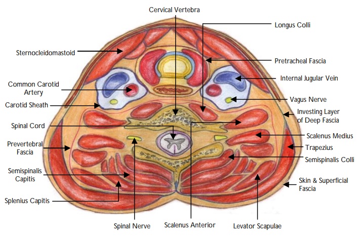
Fascial Layers of the Neck
How are the facial layers of the neck broadly divided into?
"
The facial layers of the neck are broadly divided into:
- Superficial cervical fascia
- Deep cervical fascia
Where is the superficial cervical fascia found?
"The superficial cervical fascia lies deep to the dermis and surrounds the muscles of facial expression and platysma. This layer contains fat, neurovascular bundles and lymphatics.
"
Name the different layers of the deep cervical fascia.
"
The deep cervical fascia is divided into three:
- Superficial layer (external investing layer)
- Middle layer (pre-tracheal fascia and carotid sheath)
- Internal layer or pre-vertebral fascia
Where is the superficial layer of the deep cervical fascia found?
"The superficial or investing layer of the deep fascia is the layer that surrounds the neck and wraps around the sternocleidomastoid, trapezius, muscles of mastication, submandibular and parotid glands.
"
Describe how the middle layer of the deep fascia is subdivided.
"
The middle layer of the deep fascia is subdivided into two parts: Carotid sheath and pretracheal fascia:
- Carotid sheath (vascular part) containing carotid artery (common and internal), internal jugular vein, vagus nerve and deep lymph nodes.
- Pretracheal fascia has the following components:
- Muscular part : Encloses infrahyoid muscles
- Visceral part : Encloses thyroid and parathyroid glands
- Buccopharyngeal fascia : Encompasses the pharynx and oesophagus
Describe the internal layer of deep fascia or prevertebral fascia.
The internal layer of deep fascia or prevertebral fascia is limited to the posterior neck and much thicker than the middle layer. It encloses the vertebral column and all its associated prevertebral muscles.
Identify the structures on this image.
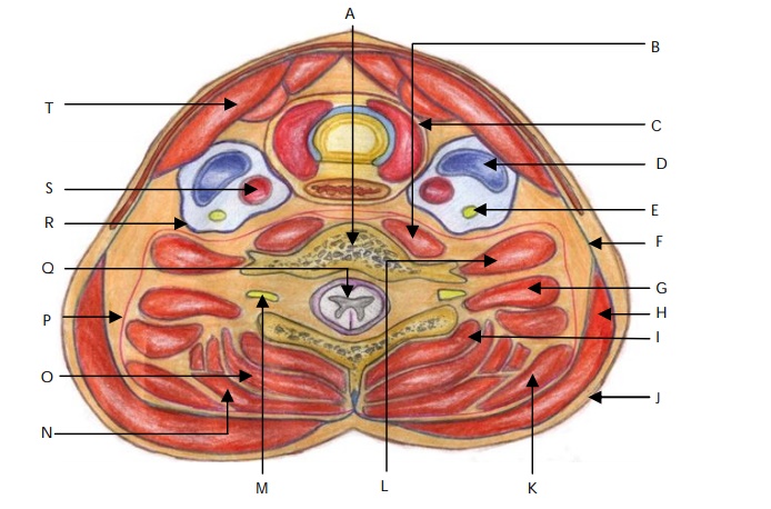
View answer
What is the role of fascia in the neck?
"
- Provides attachments to various neck muscles.
- Allows different structures to glide (larynx and trachea) or expand (pharynx and oesophagus).
- Acts as a barrier to prevent the spread of infections or malignancy.
Name the important anatomical spaces in the neck.
"
- Prevertebral space
- Pretracheal space
- Carotid space
- Danger space
- Parotid space
- Parapharyngeal space
- Retropharyngeal space
- Masseter space
- Mucosal pharyngeal space
What is the retropharyngeal space?
"The retropharyngeal space is a space posterior to pharynx and oesophagus between the pretracheal and prevertebral fascial layers.
"
What is the clinical significance of the retropharyngeal space?
"Infection of the retropharyngeal space can extend from skull to T1-T2 and can therefore result in mediastinitis or empyema.
"
What is meant by ‘the danger space’?
The ‘danger space’ lies posteriorly to the retropharyngeal space. Infections from this region may be extensions of retropharyngeal, parapharyngeal or prevertebral infections.
What is the clinical significance of the ‘danger space’?
Spread of infection within this space tends to occur rapidly due to the presence of loose areolar tissue. This can result in retropharynegeal abscess, mediastinitis,
cutaneous emphysema and sepsis.
Describe the presentation of a patient with a retropharyngeal abscess.
"This can present as difficulty swallowing, neck stiffness, neck swelling, drooling, fever and/or sepsis. The definite diagnosis is made on a CT.
"
What are the principles of management of a retropharyngeal abscess?
"
Investigations : Bloods for inflammatory markers; imaging with urgent CT scan
Treatment : High dose IV antibiotics. If required, surgical drainage can be performed via the oropharynx.
Note: In patients with significant airway obstruction secondary to oedema a surgical tracheostomy may also be indicated.
"