Name the different anatomical parts seen in a cross section of the spinal cord.
"
- Posterior horn
- Posterior funiculus
- Lateral funiculus
- Central canal
- Anterior funiculus
- Anterior horn
What is the blood supply to the spinal cord?
"
- Briefly, the spinal cord is mainly supplied by three longitudinal arteries (an anterior spinal artery and two posterior spinal arteries), along with segmental (radicular) arteries.
- These arise from the vertebral, ascending cervical, deep cervical, intercostal, lumbar and lateral sacral arteries.
- The two vertebral arteries are the first branches of the subclavian arteries.
- The ascending cervical artery is a terminal branch of the thyrocervical trunk, from the subclavian artery.
- The deep cervical arteries are also from subclavian supply, via the costocervical trunk.
- The lateral sacral arteries originate from the posterior division of the internal iliac artery.
- The paired lumbar arteries arise from the abdominal aorta and are frequently encountered during open repair of abdominal aortic aneurysms, as is the median sacral artery.
- The artery of Adamkiewicz - the great anterior segmental medullary artery - is located on the left in approximately two thirds of the population, and provides significant arterial supply to the anterior cord.
State the number of spinal nerves.
There are 31 pairs of spinal nerves, each composed of a dorsal and ventral root.
Where does the spinal cord end?
The spinal cord ends as the conus medullaris, opposite the lower end of L1 vertebrae in adults and the L3 vertebrae in children.
Name the foramen through which the spinal nerves exit the vertebral column.
Each spinal nerve exits through an intervertebral foramen.
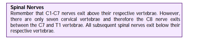
Through which vertebral level does the first cervical nerve exit the vertebral canal?
The first cervical nerve (C1) exits the vertebral canal between the skull and the first cervical vertebrae (i.e., superior to the first cervical vertebrae).
What is the functional difference between dorsal and ventral spinal roots?
Ventral roots carry, both somatic and visceral motor fibres. Dorsal roots carry only sensory fibres.
Briefly describe the function of the descending spinal tracts.
"
- The descending spinal tracts carry motor fibres via the lateral corticospinal and anterior corticospinal tracts.
- The lateral corticospinal tract is involved in fine movement of the ipsilateral limbs while the anterior corticospinal tracts supplies the central and girdle muscles.
Briefly describe the function of the ascending spinal tracts
"
- The ascending spinal tracts carry sensory fibres via the spinothalamic, dorsal column and spinocerebellar tracts
- The spinothalamic tract is responsible for pain, light touch and pressure
- The dorsal column is accountable for deep touch, proprioception and vibration sense
- The spinocerebellar tract is responsible for posture and co-ordination
"
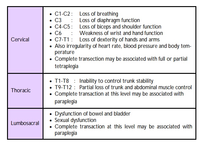
What is anterior cord syndrome?
"
- Anterior cord syndrome refers to interruption of blood flow within the anterior spinal artery supplying the anterior 2/3rd of the spinal cord.
- This may be due to trauma to the vertebra, spinal disc herniations or damage to the aorta.
- The patient will present with complete loss of muscle strength below the level of injury due to injury of the descending motor fibres in the corticospinal tract.
- Sensory loss is incomplete as ascending sensory fibres are situated in both the anterior and posterior parts of the spinal cord.
What is Brown-Sequard syndrome?
"
- Brown-Sequard syndrome refers to a loss of sensation and motor function caused by lateral hemi-section of the spinal cord.
- Can be caused by spinal cord tumours, trauma, ischaemia, infections (tuberculosis), inflammatory and autoimmune diseases.
- The patient will therefore present with spastic paralysis below the level of the lesion, ipsilateral loss of vibration, proprioception and fine touch and, contralateral loss of pain and temperature.
Name the different layers of meninges.
The cranial meninges consists of the:
Dura mater
Arachnoid mater
Pia Mater
Note: Apart from protecting the brain the meninges also enclose the cerebrospinal fluid
within the subarachnoid space (between the arachnoid and pia mater).
Name the various dural infoldings and reflections
The internal meningeal layer of the dura form dural infoldings, which separate the regions of the brain from each other:
• Cerebral falx - also known as Falx cerebri - largest dural reflection, lies within the longitudinal fissure and separates the right and left cerebral hemispheres.
• Cerebellar tentorium - also known as Tentorium cerebelli - second largest reflection separating the occipital lobes of the cerebral hemispheres from the cerebellum.
• Cerebellar falx - also known as Falx cerebelli - lies inferior the cerebellar tentorium and partially separates the cerebellar hemispheres.
• Sellar diaphragm - also known as Diaphragma sellae - smallest dural folding and covers the pituitary gland.
Describe the ganglia in the head and neck region.
"
There are four main ganglia, all parasympathetic, in the head and neck region. They are as follows:
- Ciliary ganglion: Parasympathetic ganglion located behind the eye in the posterior orbit, associated with the occulomotor nerve and contributes to the pupillary and accommodation reflexes.
- Pterygoplatatine ganglion: Parasympathetic ganglion largely associated with the greater petrosal nerve (branch of facial nerve) supplying the lacrimal gland, paranasal sinuses and glands in the palate and nasal cavity.
- Submandibular ganglion: Parasympathetic ganglion associated with the chorda tympani branch of the facial nerve and lingual branch of V3 supplying the submandibular and sublingual glands.
- Otic ganglion: Parasympathetic ganglion located just below the foramen ovale in the infratemporal fossa associated with the glossopharyngeal nerve secretomotor to the parotid gland.
"