Describe the indications for intercostal chest drain insertion.
"
Some indications for insertion of an intercostal chest drain include are as follows:
- Tension pneumothorax following needle decompression.
- Traumatic haemopneumothorax.
- Pneumothorax in a ventilated patient.
- Persistent or recurrent pneumothorax following aspiration.
- Postoperative e.g., following thoracic surgery.
- Malignant pleural effusion.
- Empyema and complicated para-pneumonic pleural effusion.
What is a tension pneumothorax?
"A tension pneumothorax is when air collects within the pleural space and cannot escape, causing shift of the mediastinal structures, either through a breach of the chest wall or airway.
"
What are the consequences of a tension pneumothorax?
"In a tension pneumothorax, air is unable to escape from the pleural space causing mediastinal shift leading to pressure on large veins and a subsequent reduction in cardiac output.
"
What are the anatomical landmarks for needle decompression of a tension pneumothorax?
"For immediate decompression of a tension pneumothorax, a large bore needle should be inserted into the second intercostal space, just above the third rib (since the neurovascular bundle lies just below the rib in the costal groove), in the midclavicular line of the involved hemithorax.
"
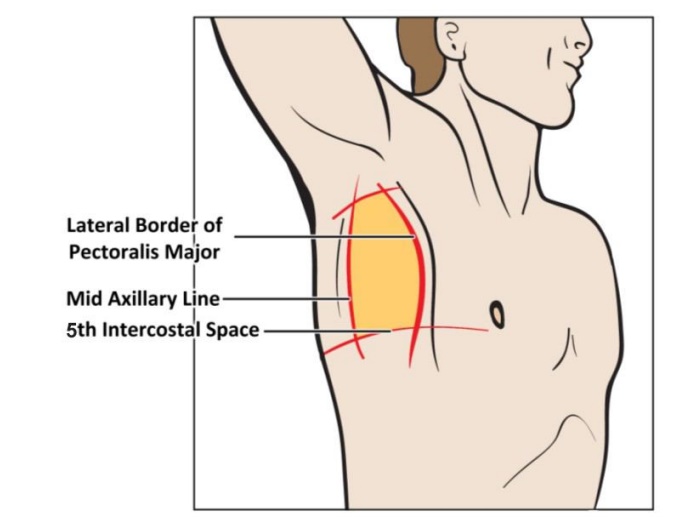
Describe the anatomical landmarks for the safe insertion of an intercostal chest drain.
The anatomical landmarks for intercostal chest drain insertion describe the ‘triangle of safety’:
Anterior : Posterior (lateral) border of pectoralis major.
Posterior : Mid-axillary line.
Apex : Axilla.
Base : A horizontal line drawn along the upper surface of the 6th rib.Within this triangle of safety, the intercostal drain can be inserted in the 5th intercostal space, above the 6th rib, just anterior to the mid-axillary line to avoid damage to the long thoracic nerve (this nerve runs in the mid-axillary line).
Name the layers you would go through when inserting an intercostal chest drain.
"
When inserting a intercostal drain, the structures one would go through from superficial to deep are the:
- Skin.
- Subcutaneous fat.
- External intercostal muscles (infero-anterior orientation of muscle fibres).
- Internal intercostal muscles (infero-posterior orientation of muscle fibres).
- Innermost intercostal muscles (like internal intercostal; only found at the lateral most part of the intercostal space).
- Parietal pleura.
Describe the arterial supply to the chest wall.
"
The arteries supplying the chest wall arise from the thoracic aorta, subclavian artery and axillary arteries:
Posterior intercostal arteries: Arising from the superior intercostal artery (supplies intercostal spaces 1 and 2) which (a branch of the costocervical trunk arising from the subclavian artery). The rest of the lower 9 intercostal spaces are supplied by direct branches from the descending thoracic aorta.
- Anterior intercostal arteries: Arising from the internal thoracic (intercostal spaces 1-6) and musculophrenic arteries (intercostal spaces 7-9).
- Superior and lateral thoracic arteries: Arising from the axillary artery, suppies the anterior and lateral chest wall.
- Subcostal arteries: Arising from the descending thoracic aorta, these arteries supply the areas beneath the 12th rib and the anterolateral chest/abdominal wall.
Describe the nerve supply to the chest wall.
"
The chest wall is supplied by 12 pairs of thoracic spinal nerves which divide into ventral and dorsal rami:
- Ventral rami: Ventral rami of T1-T11 form the intercostal nerves that run in the intercostal spaces; T12 forms the subcostal nerves.
- Dorsal rami: Run postero-lateral to the vertebrae to supply bones, joints, deep back muscles and skin of thoracic region.
Describe the venous drainage of the chest well.
"
- Veins accompany arteries and nerves, and lie most superior in the costal grooves.
- The anterior aspect of the thorax is drained by the anterior intercostal veins, which in turn drain into the internal thoracic vein and then the brachiocephalic vein.
- The posterior aspect of the thorax is drained by the azygos system into the SVC:
- The 1st intercostal space on both sides is drained by the highest (supreme) intercostal vein and then into the brachiocephalic vein.
- The 2nd, 3rd and 4th posterior intercostal veins drain into the superior intercostal vein. On the right side, the superior intercostal vein drains into azygos vein. On the left side, it drains into the accessory hemiazygos vein or directly to the brachiocephalic vein.
- The rest of the 8 lower posterior intercostal spaces are drained by the azygos venous system, which empties into the SVC. On the left side, intercostal spaces 5-8 are drained by the accessory hemiazygos vein and intercostal spaces 9-12 are drained by the hemiazygos vein. The accessory hemiazygos and hemiazygos veins drain into the azygos system before it empties into the SVC.