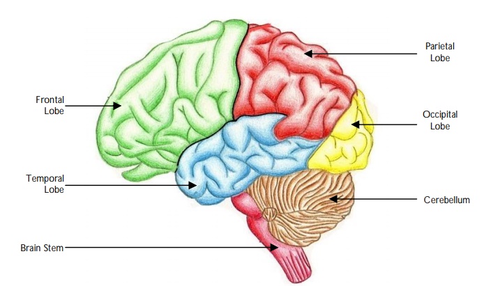
Identify the following parts of the brain on the image
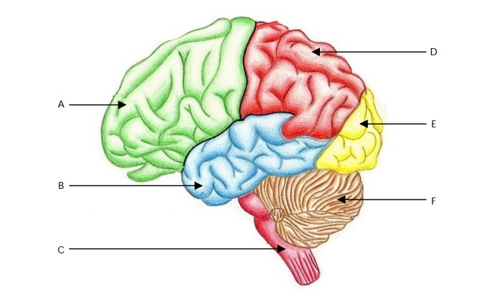
View answer
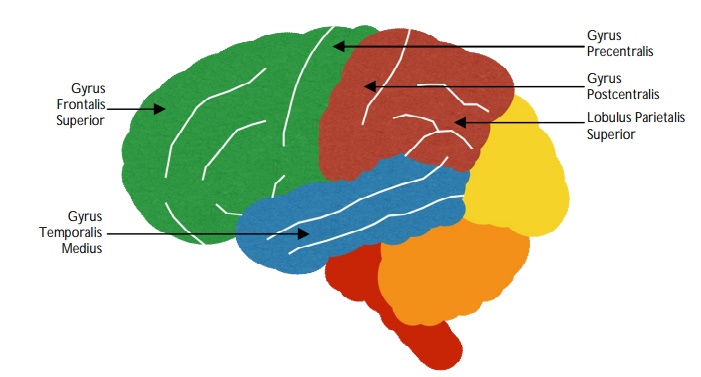
Identify the following parts of the brain on the image
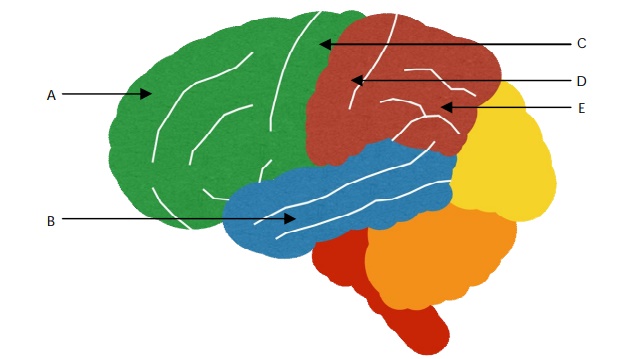
View answer
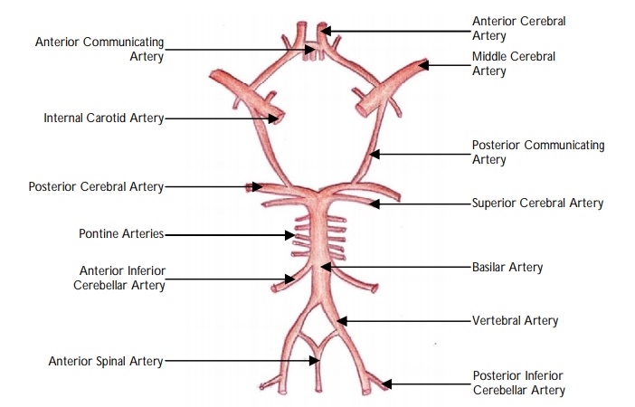
Identify the following arteries on the image.
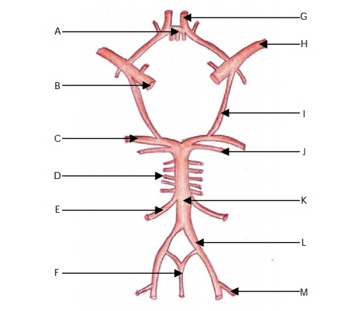
View answer
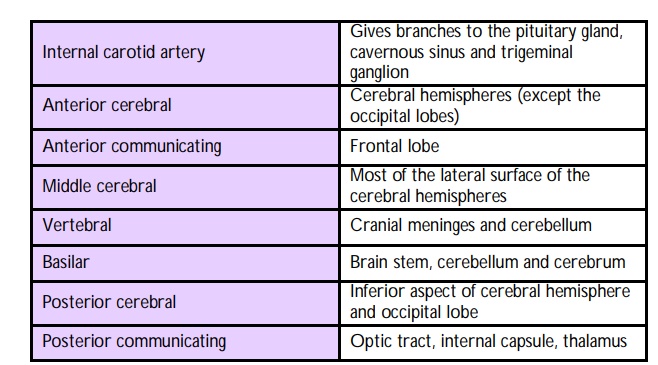
Describe the blood supply to the brain.
The brain derives its blood supply from the internal carotid and the vertebral arteries.
The terminal branches of the internal carotid arteries are the anterior and middle cerebral arteries. The two anterior cerebral arteries are connected together via the anterior communicating arteries.
The vertebral arteries arise in the root of the neck, ascend cranially and unite at the caudal border of the pons to form the basilar artery. The basilar artery ascends and divides into the two posterior cerebral arteries. The later join the termination of the internal carotid arteries to form the circle of Willis.
Describe the venous drainage of the cerebrum.
"
The venous drainage of the cerebrum is by the superficial and deep systems. The superficial system, composed of dural venous sinuses (the most important is the superior saggital sinus), form the confluence of sinuses. From here, two transverse sinuses bifurcate into the sigmoid sinuses which form the two jugular veins.
The deep venous drainage system joins behind the midbrain to form the vein of Galen. This vein merges with the inferior saggital sinus to form the straight sinus and joins the superficial venous system.
"
Name the venous sinuses of the brain.
"
- Superior sagittal sinus
- Inferior sagittal sinus
- Straight sinus
- Transverse sinus
- Confluence of sinuses
- Sigmoid sinus
- Occipital sinus
- Cavernous sinus
- Superior petrosal sinus
- Inferior petrosal sinus