LOWER LIMB

OSCE
Station 4

Gluteal Region

"
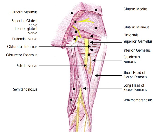
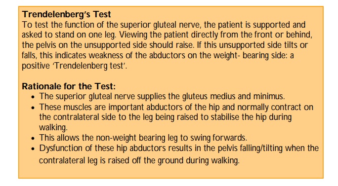
  • Gluteus minimus and medius: Abduct and medially rotate the thigh. They play an important role during walking to stabilise the unsupported side of the pelvis to prevent it from tilting during walking.
  • Gluteus maximus: Extends and externally rotates the thigh and, abducts the hip. It supports the extended knee through the iliotibial tract and has a key role in functions such as standing from a sitting position, walking up the stairs and running.

Note: A deeper group of smaller muscles (piriformis, obturator internus and externus, gemelli superior and inferior and quadratus femoris) are covered by the inferior half of gluteus maximus and are the lateral rotators of the thigh. They also help to stabilise the hip joint.

"

"

  • Gluteus maximus :        Inferior gluteal nerve (S1 and S2).
  • Gluteus medius and gluteus minimus   :        Superior gluteal nerve (L5 and S1).

"

"The superior and inferior gluteal arteries originate from the internal iliac artery. The superior gluteal artery is the largest branch of the internal iliac and a continuation of the posterior division of this vessel. The inferior gluteal artery is one of the two terminal branches of the anterior division of the internal iliac artery (the other terminal branch is the internal pudendal artery). "
"

Superior gluteal artery:

  • Passes posteriorly between the lumbosacral trunk and the 1st sacral nerve.
  • Leaves the pelvis through the greater sciatic foramen, superior to the piriformis.
  • Divides immediately into superficial and deep branches.
  • The superficial branch supplies the gluteus maximus and skin overlying its proximal attachment.
  • The deep branch supplies the gluteus medius, gluteus minimus and tensor fascia lata.
  • It anastomoses with the inferior gluteal and medial circumflex femoral vessels.


Inferior gluteal artery:

  • Passes posteriorly through the parietal pelvic fascia, between the 1st and 2nd sacral nerves.
  • Exits the pelvis through the greater sciatic foramen, inferior to the piriformis.
  • Enters the gluteal region deep to gluteus maximus, and descends medial to the sciatic nerve.
  • Supplies the gluteus maximus, obturator internus, quadratus femoris and superior parts of the hamstrings.
  • It anastomoses with the superior gluteal artery and contributes to the cruciate anastomosis of the thigh.
"

Identify the points on the image:
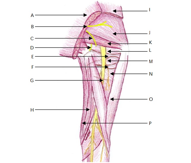
"The surface marking of the superior border of the piriformis is indicated by a line joining the skin dimple formed by the posterior superior iliac spine to the superior border of the greater trochanter of the femur. "
"

The sacral plexus is formed from the anterior primary rami of L4–L5, S1–S4.

Note: The sacral nerves (S1-S4) emerge from the anterior sacral foramina and unite in front of piriformis where they are joined by the lumbar trunk (L4, 5) to form the lumbosacral trunk.

"
"
  • Antero-laterally :           Greater sciatic notch of the ilium.
  • Postero-medially :           Sacrotuberous ligament.
  • Inferiorly :           Sacrospinous ligament and the ischial spine.
  • Superiorly :           Anterior sacroiliac ligament.
"
"

The structures passing through the greater sciatic foramen above the piriformis are:

  • Superior gluteal vessels.
  • Superior gluteal nerve.
"
"

Structures passing through the greater sciatic foramen below the piriformis

  • Inferior gluteal vessels.
  • Inferior gluteal nerve.
  • Internal pudendal vessels.
  • Pudendal nerve.
  • Sciatic nerve.
  • Posterior cutaneous nerve of the thigh.
  • Nerve to quadratus femoris.
  • Nerve to obturator internus.
"
"
  • Anterior :            Tuberosity of the ischium.
  • Superior :            Spine of the ischium and sacrospinous ligament.
  • Posterior :            Sacrotuberous ligament.
"

Piriformis : Branches of ventral rami of S1 and S2.
Obturator externus : Obturator nerve L3 and L4.
Obturator internus : Nerve to obturator internus L5 and S1.
Superior gemellus : Nerve to obturator internus L5 and S1.
Inferior gemellus : Nerve to quadratus femoris L5 and S1.
Quadratus femoris : Nerve to quadratus femoris L5 and S1.

"

To surface mark the sciatic nerve:

  • Identify the posterior superior iliac spine, greater trochanter and ischial tuberosity.
  • Draw a curved line half way between the PSIS and the greater trochanter and continue the line half way between the greater trochanter and ischial tuberosity.
  • The nerve runs in the midline of the posterior aspect of the thigh between the hamstrings towards the popliteal fossa.
  • The two divisions of the nerve, the tibial and common peroneal nerves, can divide anywhere along the course but most commonly approximately 5-7 cm above the posterior knee joint line.
"
"Intramuscular injections should be performed in the upper outer quadrant of the buttock in order to avoid iatrogenic injury to the sciatic nerve. "
"

The sciatic nerve usually passes beneath the piriformis in approximately 85-90% of the population. Other variations include:

  • Piriformis divided into two parts with the peroneal division of the sciatic nerve passing between the two parts of piriformis.
  • The peroneal division of the sciatic nerve passes over the piriformis and the tibial division passes beneath the undivided muscle.
  • In these cases the entire nerve passes through the divided piriformis.
"