LOWER LIMB
OSCE
Station 3
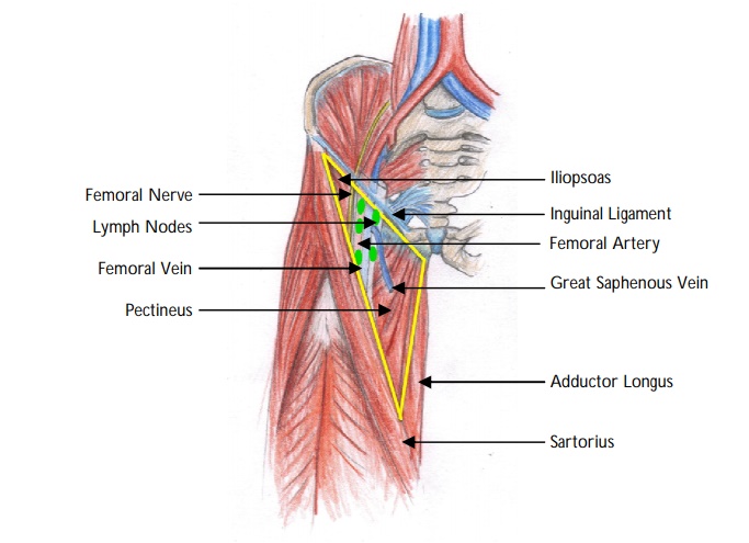
Femoral Triangle
Name the boundaries of the femoral triangle.
"
- Lateral : Medial border of sartorius.
- Medial : Medial border of adductor longus.
- Superior : Inguinal ligament.
- Floor : Iliacus, psoas major and pectineus.
- Roof : Skin, subcutaneous tissue and fascia lata.
"
Name the contents of the femoral triangle.
"
The contents of the femoral triangle (from lateral to medial) are:
- Femoral nerve and its branches.
- Femoral artery and several of its branches.
- Femoral vein and its proximal tributaries.
- Femoral canal and lymph nodes.
Identify the points on the image
View answer
What are the origins and insertions of iliacus and psoas major muscles?
"
- The psoas major originates along the lateral surfaces of the vertebral bodies of T12 and L1-L5 and their associated intervertebral discs.
- The iliacus originates in the iliac fossa of the pelvis.
- Psoas major unites with the iliacus at the level of the inguinal ligament and crosses the hip joint to insert on the lesser trochanter of the femur.
What is the action of the iliopsoas muscle?
"The iliopsoas acts conjointly to flex and laterally rotate the hip joint. If the limb is fixed, it helps in flexion of the trunk.
"
Where is the mid-inguinal point and what is the clinical significance?
"
- The mid-inguinal point is halfway between the pubic symphysis and the anterior superior iliac spine.
- The femoral artery crosses into the lower limb and can be palpated at this anatomical landmark.
What is the femoral sheath?
| The femoral sheath is a shaped fascial tube, the femoral sheath, extends 3-4 cm inferior to the inguinal ligament and encloses the proximal parts of the femoral vessels and the femoral canal. It is a continuation of the transversalis fascia anteriorly and the iliopsoas fascia posteriorly. The sheath ends by becoming continuous with the adventitia covering the femoral vessels. It allows the femoral vessels to glide deep to the inguinal ligament during movement of the hip. |
Name the contents of the femoral sheath.
| The femoral sheath encloses the proximal parts of the femoral artery, femoral vein and femoral canal. The femoral sheath does not enclose the femoral nerve. |
What is the femoral canal?
| The femoral sheath is subdivided into three compartments (lateral, intermediate and medial) by vertical septa which are derived from extra-peritoneal connective tissue of the abdomen that extends along the vessels. The femoral canal is the medial compartment of the femoral sheath. The femoral canal allows the femoral vein to expand during increased venous return of the lower limb. |
What are the contents of the femoral canal?
| The femoral canal contains fat, lymphatics and Cloquet’s node. |
What is the clinical significance of Cloquet’s node?
| Cloquet’s node drains the lower limb, perineum and anterior abdominal inferior to the umbilicus. It may be enlarged in cases of carcinoma or infection at these sites. |
What is the femoral ring?
The base of the femoral canal (the abdominal end) is directed superiorly and, although oval-shaped, is called the ‘femoral ring’.
Name the boundaries of the femoral ring
Anterior : Inguinal ligament.
Posterior : Pectineal ligament.
Medial : Lacunar ligament.
Lateral : Femoral vein.
What is the clinical significance of the femoral ring.
| Femoral hernias can enter the thigh through the femoral ring. They may present as a tender mass in the femoral triangle, infero-lateral to the pubic tubercle. The hernia is bounded by the femoral vein laterally and the lacunar ligament medially. |
Describe the course of the femoral artery.
- The external iliac artery becomes the common femoral artery at the level of the inguinal ligament (mid-inguinal point).
- Approximately 4 cm into the femoral triangle, the common femoral artery divides into the deep profunda femoris artery and the superficial femoral artery.
- The profunda femoris artery gives off the medial and lateral circumflex femoral arteries.
- The superficial femoral artery runs to the apex of the femoral triangle, enters the adductor canal and exits the adductor hiatus to form the popliteal artery.