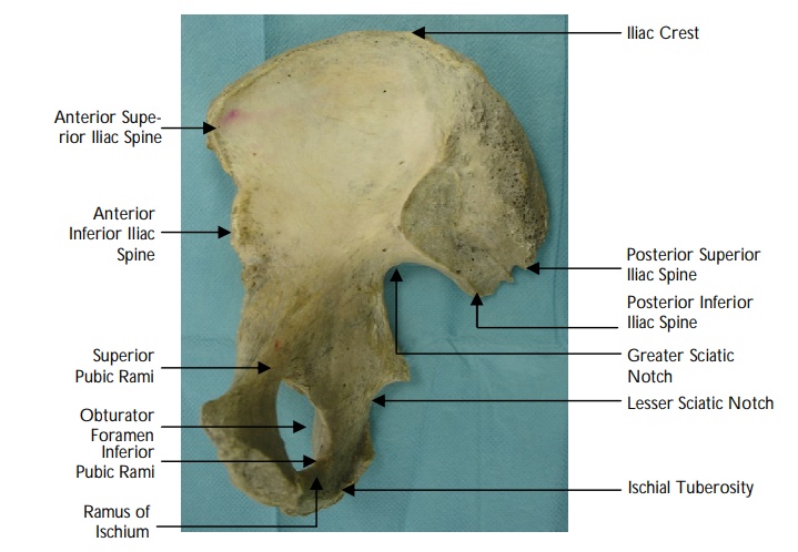
Identify the points on the image
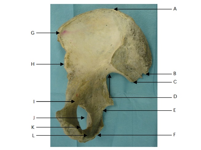
View answer
Name the extracapsular ligaments of the hip capsule and their attachments.
"
The extracapsular ligaments are the:
- Ilio-femoral (Y-shaped and ‘twisted’): Arises from the anterior inferior iliac spine, bifurcates, and is inserted at each end of the trochanteric line (resists hyperextension).
- Pubo-femoral: Arises from the ilio-pubic junction to blend with the medial aspect of the capsule (resists extension and abduction).
- Ischio-femoral: Arises from the ischium and inserts into the base of the greater trochanter (limits extension).
Note: All three strengthen the capsule and prevent an excessive range of movement of the hip joint. The ilio-femoral is the strongest of the hip ligaments (and one of the strongest ligaments in the body).
"
Name the muscles which attach to the greater trochanter of the femur.
"
The greater trochanter is the distal insertion of the following muscles:
- Lateral surface : Gluteus medius.
- Anterior surface : Gluteus minimus.
- Superior border : Piriformis.
- Postero-medial surface : Gemelli superior and inferior and, obturator internus.
Describe the blood supply to the hip joint.
"
- The hip joint is supplied by two important anastomoses- the cruciate and trochanteric anastomoses. These two anastomoses connect the femoral artery or profunda femoris to the gluteal vessels.
- The cruciate anastomoses is formed by the inferior gluteal artery, the lateral and medial circumflex femoral arteries, the first perforating artery from profunda femoris and the posterior branch of the obturator artery.
- The trochanteric anastomosis comprises of the superior gluteal artery and the medial and lateral superior circumflex arteries.
Describe the blood supply to the neck and head of the femur.
"
The blood supply to the neck and head of femur can be broadly grouped into:
- The retinacular arteries: The femoral artery gives off the deep profunda femoris branch which divides into medial and lateral circumflex femoral arteries. The lateral circumflex femoral artery gives off the transverse branch which further divides into ascending and descending branches. The ascending branch (also known as the ascending cervical arteries) contribute to the sub-synovial arterial ring and travels proximally under the hip capsule, deep to the synovial membrane, towards the femoral head. The medial circumflex femoral artery, located on the posterior-superior aspect of the femoral neck, is the main blood supply for the weight-bearing dome of the femoral head and therefore is significant.
- Artery of ligamentum teres: The obturator artery contributes to the epiphyseal arteries and the artery of ligamentum teres which supplies a variable proportion of the femoral head. The artery of ligamentum teres can also arise from the medial circumflex femoral artery.
- Interosseous blood supply: The intramedullary branches of nutrient artery, metaphyseal, and epiphyseal vessels supply both the marrow and the cortex.
- Extracapsular ring of anastomosis: Ascending cervical vessels also contribute to the extracapsular ring of anastomosis at the base of the femoral neck.
- Perforating arteries: From the profunda femoris supply the shaft of the femur.
What is the clinical relevance of this blood supply?
"
- Intracapsular fractures of the neck of femur disrupt retinacular vessels and compromise the blood supply to the femoral head.
- Rupture of these vessels caused by a fracture can result in avascular necrosis of femoral head as the blood supply through the ligamentum teres is usually inadequate in adults.
"
What is the innervation of the hip joint?
"The hip joint is innervated by the sciatic, femoral and obturator nerves.
"
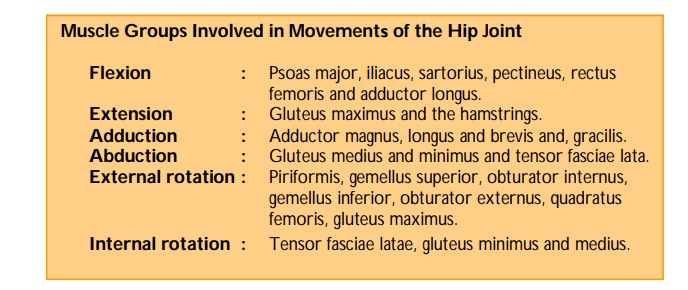
Name the main flexor muscles of the hip.
"Psoas major and iliacus muscles are the main hip flexors. Other muscles which contribute include pectineus, rectus femoris, adductor longus and sartorius.
"
What is the nerve supply to the main flexor muscles of the hip?
"
- Psoas major is supplied by the ventral rami of L1, L2 spinal nerves, with a small contribution from L3.
- Iliacus, rectus femoris and sartorius are innervated by the femoral nerve (L2-4).
- Pectineus is supplied by the femoral nerve in the majority of cases. However in 20% of the population it is supplied by the accessory obturator nerve (a branch of the obturator nerve).
Name the external rotators of the hip.
"
The external rotators of the hip are the:
- Piriformis.
- Gemellus superior and inferior.
- Obturator externus and internus.
- Quadratus Femoris.
- Gluteus Maximus.
Note: The superior and inferior gemelli join together along with the obturator internus to form the conjoined tendon.
"
What are the different approaches for surgical exposure of the hip joint?
The lateral approach.
The anterior approach.
The posterior approach.
Briefly describe the different surgical approaches to the hip joint.
Lateral approach:
Involves dividing the fibres of tensor fasciae latae, gluteus medius and minimus to expose the femoral neck. Further access can be obtained by detaching the greater trochanter from its gluteal insertions.
Anterior approach:
The incision is made between gluteus medius and minimus laterally and sartorius medially. The head of rectus femoris is divided and reflected to expose the anterior aspect of the hip joint. Further access can be obtained by detaching gluteus medius and minimus from the ilium.
Posterior approach:
The incision is placed in a curvilinear fashion over the posterior superior iliac spine to the greater trochanter and then continues
vertically downwards from this point.
Name the structures at risk of injury during the posterior approach.
"The sciatic nerve and superior gluteal nerve are at risk during the posterior approach to the hip joint.
"