UPPER LIMB
OSCE
Station 4
Antecubital Fossa
Name the boundaries of the antecubital fossa.
"
- Superior : Imaginary line connecting the medial and lateral humoral epicondyles.
- Medial : Lateral border of pronator teres.
- Lateral : Medial border of brachioradialis.
- Roof : Skin/ subcutaneous tissue and fascia, reinforced by the bicipital aponeurosis.
- Floor : Brachialis medially and supinator laterally.
Identify the labeled structures on this image
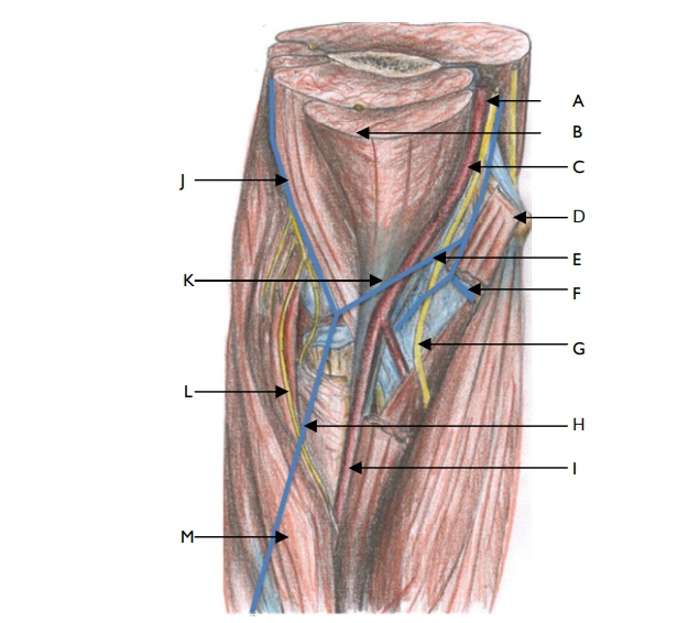
View answer
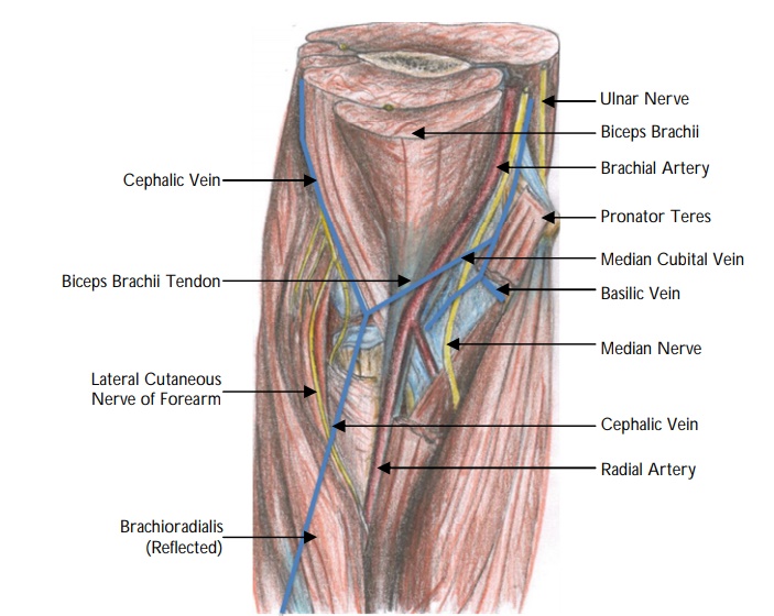
Name the contents of the antecubital fossa.
"
The contents of the antecubital fossa (from radial to ulnar) are the:
- Biceps brachii Tendon.
- Brachial Artery (terminal part).
- Median Nerve.
"Name the structures which lie on the surface of the cubital fossa. "
"
The structures which lie on the surface of the cubital fossa from medial to lateral are the:
- Median basilic vein.
- Median antebrachial vein.
- Median cutaneous nerve of the forearm.
- Median cephalic vein.
- Lateral cutaneous nerve of the forearm.
"
Why is an understanding of the anatomy of the cubital veins clinically significant?
"
- The cubital fossa is a common site for venepuncture and intravenous injection due to the prominence and accessibility of these veins.
- They can easily be seen and accessed when a tourniquet is applied to the arm.
- There is considerable variation in the pattern of veins in the cubital fossa. In approximately 20% of the population the median antebrachial vein divides into a median basilic vein and a median cephalic vein – in these cases a clear ‘M’ formation is produced by the cubital veins. These veins are ideal sites for venepuncture and are most commonly used.
- The cubital veins are also used for the introduction of cardiac catheters to the right side of the heart for monitoring of intra-cardiac pressures and for blood sampling.
Name the boundaries of the cubital tunnel.
"
- Roof : Arcade of fibres from the medial epicondyle of the humerus to the olecranon of the ulna formed by the aponeurotic expansion of the two heads of the flexor carpi ulnaris (Osborne ligament).
- Floor : Formed by the medial collateral ligament of the elbow which attaches from the medial border of the olecranon to the base of the medial epicondyle.
What is cubital tunnel syndrome?
"Cubital tunnel syndrome is compression of the ulnar nerve at the elbow as it runs through the cubital tunnel.
"
Describe the clinical symptoms and signs of cubital tunnel syndrome.
"
- Symptoms : Pain, numbness, weak grip.
- Signs : Tinel’s at the medial elbow; Froment’s sign (flexion of the thumb interphalangeal joint with attempting to adduct the thumb towards the index finger); Wartenberg’s sign (involuntary abduction of the little finger caused by unopposed action of the extensor digiti minimi); sensory disturbance of the ring and little finger; intrinsic muscle wasting.
Note: Wartenberg’s sign should not be confused with Wartenberg’s syndrome, which is compression of the superficial branch of the radial nerve at the wrist presenting with paraesthesia in the first dorsal webspace.
"
What is the management of cubital tunnel syndrome?
"
- Conservative : Splinting, therapy.
- Surgical decompression : Of the ulnar nerve at the level of the cubital tunnel (can be performed alone or with transposition of the ulnar nerve).
Note: Surgical decompression involves release of the Osborne ligament and the arcade of Struthers.
"
Name some other common sites of ulnar nerve compression.
"
- Arcade of Struthers (musculofascial band approximately 8 cm proximal to the medial epicondyle).
- Medial intermuscular septum.
- Olecranon groove.
- Flexor-pronator aponeurosis (as the nerve exits the flexor carpi ulnaris, it perforates a fascial layer between the flexor digitorum superficialis and the flexor digitorum profundus).
- Guyon’s canal.
"