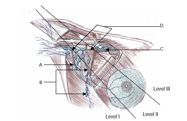
Label the lymph node groups on the image
View answer
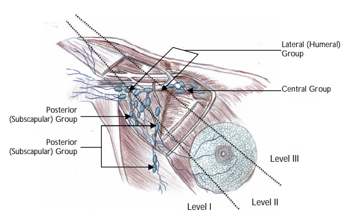
Why are the levels of axillary nodes clinically significant in breast cancer?
The levels of axillary lymph nodes are particularly significant in the staging of breast cancer and axillary node surgery (axillary sampling and axillary node clearance). Level III is the most aggressive type of dissection as it removes all of the nodal tissue from the axilla.
Note: The axillary nodes can also be involved in skin cancers such as malignant melanoma and squamous cell carcinoma.
Name the structures that can be damaged during axillary node clearance surgery.
Axillary vein.
Long thoracic nerve.
Thoracodorsal nerve.
Intercostobrachial nerve (union of the lateral cutaneous branch of the second intercostal nerve with a filament from the medial cutaneous nerve of the arm).
How do injuries to these nerves clinically present?
Division or damage to the long thoracic nerve (that supplies serratus
anterior) presents with winging of the scapula.
Division or damage to the thoracodorsal nerve (that supplies the latissimus dorsi [LD]) presents with weakness of medial rotation and adduction of the arm. Patients will find it difficult to perform activities such as climbing and swimming that require action of the LD muscle.
Note: The LD muscle can also be used for reconstruction of the breast following mastectomy therefore, it is particularly important to preserve the blood supply and innervation to this muscle in these patients.
The intercostabrachial nerve innervates the skin of the upper half of the medial and posterior part of the arm. Division or damage to this nerve results in loss of sensation to this area.
Where would you palpate the axillary artery in the axilla?
The axillary artery can be palpated in the inferior part of the lateral wall of the axilla.
Note: If there is profuse bleeding from the axilla (such as following trauma i.e., a bullet or stab wound to the axilla) compression of the third part of the axillary artery against the humerus can be attempted to control the bleeding. In a more proximal injury, it can also be compressed at its origin (as the subclavian artery crosses the first rib) by putting downward pressure between the clavicle and attachment of the sternocleidomastoid.
Name the branches of the axillary artery.
The axillary artery is divided into three parts which are named by their relation to
pectoralis minor:
First Part (medial to pectoralis minor) has one branch:
• Superior thoracic artery.
Second Part (behind pectoralis minor) has two branches:
• Thoracoacromial trunk.
• Lateral thoracic artery.
Third Part (lateral to pectoralis minor) has three branches:
• Subscapular artery.
• Anterior circumflex humeral artery.
• Posterior circumflex humeral artery.
Briefly describe the course of the axillary nerve.
The axillary nerve is the terminal branch of the posterior cord of the brachial plexus receiving fibres from C5 and C6. It passes to the posterior aspect of the arm, supplying teres minor as it exits the axilla through the quadrangular space. It then supplies deltoid from its posterior aspect and winds around the surgical neck of the humerus. It continues as the superior lateral brachial cutaneous nerve which innervates the skin.
What are the boundaries of the quadrangular space?
Superior : Subscapularis and teres minor.
Inferior : Teres major.
Medial : Long head of triceps.
Lateral : Humerus.
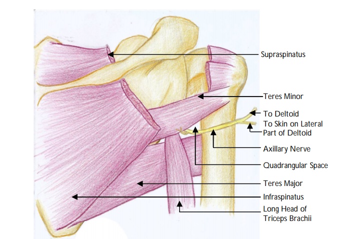
Name the other structure that passes through the quadrangular space.
The posterior circumflex humeral artery also traverses the quadrangular space
What is the clavipectoral fascia?
The clavipectoral fascia extends from the axillary fascia, encloses the pectoralis minor and subclavius then attaches superiorly to the clavicle. The clavipectoral fascia superior to pectoralis major (the costocoracoid membrane) is pierced by the lateral pectoral nerve. The part of the clavipectoral fascia inferior to pectoralis minor is the suspensory ligament of the axilla.
Name the structures that pass through the clavipectoral fascia.
The structures passing through the clavipectoral fascia include the:
Lateral pectoral nerve.
Thoracodorsal trunk.
Cephalic vein.
Lymphatics
What is the triangular interval and what are its boundaries?
The triangular interval is a space found in the axilla. It is bounded by:
Superior : Teres major.
Medial : Long head of triceps.
Lateral : Humerus.
Note: The triangular interval is also referred to as the lateral triangular space, lower
triangular space, and triceps hiatus
What are the contents of the triangular interval?
The radial nerve and profunda brachii artery pass through the triangular interval to
the posterior compartment of the arm.