Describe the structure of the diaphragm.
"
- The diaphragm is a dome-shaped musculo-fibrous structure that separates the thoracic and abdominal cavities.
- It consists of two parts: the peripheral muscle and central tendon.
- The peripheral muscle is made up of many radial muscle fibres originating on the ribs, sternum, and spine that converge on the central tendon.
- The central tendon is a flat aponeurosis made of dense collagen fibres that acts as the tough insertion point of the muscles.
- The peripheral muscle can be further divided by its origins into the sternal, costal and lumbar regions:
- The sternal region is made up of two small muscular segments that attach to the posterior aspect of the xiphoid process.
- The costal region is made up of several wide muscle segments whose origins are found on the internal surface of the inferior six ribs and costal cartilages.
- The lumbar part, arising from the medial and lateral arcuate ligaments and the three superior lumbar vertebrae. This lumbar part forms the right and left crura. The medial arcuate ligament is formed from the psoas major fascia, and the lateral from the fascia covering quadratus lumborum and lateral arcuate ligaments.
What is the arterial supply to the diaphragm?
"
- The superior surface of the diaphragm is supplied by the pericardiophrenic artery and musculophrenic arteries from the internal thoracic artery, superior phrenic arteries from the descending thoracic aorta and the lower internal intercostal arteries.
- The inferior surface of the diaphragm is supplied by the inferior phrenic arteries from the abdominal aorta.
Describe the venous and lymphatic drainage of the diaphragm.
"
Superior surface:
- Venous : Musculophrenic and pericardiophrenic veins drain into internal thoracic veins, superior phrenic vein drains into IVC.
- Lymphatic : Diaphragmatic nodes to phrenic, parasternal and posterior mediastinal lymph nodes.
Inferior surface:
- Venous : Inferior phrenic veins (draining into IVC) and suprarenal vein.
- Lymphatic : Superior lumbar lymph nodes.
What is the innervation of the diaphragm?
"
The innervation of the diaphragm is:
- Motor supply : Phrenic nerves C3-C5.
- Sensory supply : Phrenic nerves C3-C5 (central), intercostal T5-T11 (peripheral) and T12 (subcostal)
Name the three major openings in the diaphragm.
The major openings in the diaphragm are:
T8 : Caval opening.
T10 : Oesophageal hiatus.
T12 : Aortic hiatus.
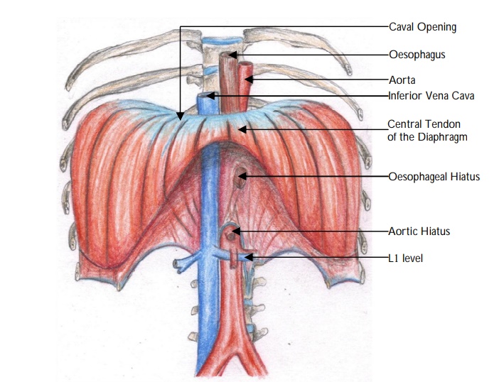
Where is the caval opening located?
"The caval opening is located in the central tendon.
"
What structures pass through the caval opening?
"The inferior vena cava and right phrenic nerve pass through the caval opening.
"
Where is the oespophageal hiatus located?
"
The oesophageal hiatus is found in the right crus.
Note: The right crus, larger and longer than the left crus, arises from the first 3-4 lumbar vertebra; the left crus arises from the first 2-3 lumbar vertebra.
"
What structures pass through the oesophageal hiatus?
"The oesophagus, posterior and anterior vagal trunks and oesophageal branch of left gastric artery pass through the oesophageal hiatus.
"
How is the aortic hiatus formed?
"The aortic hiatus is formed by the right and left crura and the median arcuate ligament.
"
What structures pass through the aortic hiatus?
"The aorta, azygos vein and thoracic duct pass through the aortic hiatus.
"
What other structures traverse the diaphragm?
The other structures traversing the diaphragm include the:
Greater and lesser right and left splanchnic nerves (pass through the aperture of right and left crus)
Sympathetic trunk (passes behind the diaphragm behind the medial lumbocostal arch).
Superior epigastric branch of internal thoracic artery (passes through the areolar tissue between the sternal and costal parts).
The left phrenic nerve passes through its own hiatus at the periphery of the diaphragm.
Which structures may be injured during surgery to the oesopheageal hiatus?
During surgery to the oesophageal hiatus the structures which also pass through the hiatus may be injured. These include the vagal trunks, left inferior phrenic 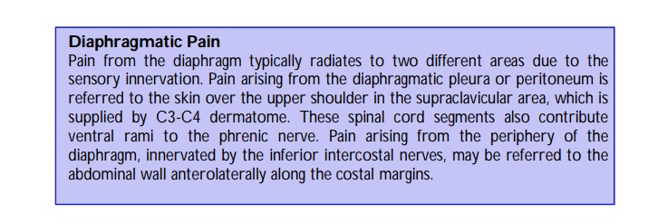 vessels, oesophageal branches of the left gastric vessels.