Name the parts of the cranial fossae.
"
The cranial fossae is divided into three parts:
- Anterior
- Middle
- Posterior
Describe the bony boundaries of the anterior cranial fossa.
"
The boundaries of the anterior cranial fossa are:
- Anterior : Inner surface of frontal bones
- Posterior-medial : Anterior border of the prechiasmatic sulcus of sphenoid (also called limbus)
- Posterior-lateral : Lesser wings of sphenoid
- Floor : Orbital part of frontal bone, ethmoid bone and the anterior aspects of the body and lesser wings of the sphenoid
Describe the bony boundaries of the middle cranial fossa.
"
The boundaries of the middle cranial fossa are:
- Anterior-lateral : Lesser wings of sphenoid
- Anterior-medial : Chiasmatic sulcus and limbus of sphenoid
- Posterior-lateral : Petrous part of temporal bone
- Posterior-medial : Dorsum sellae of the sphenoid
- Floor : Body and greater wing of the sphenoid; squamous and petrous parts of the temporal bone.
Describe the bony boundaries of the posterior cranial fossa.
"
The boundaries of the posterior cranial fossa are:
- Anterior-medial : Dorsum sellae of the sphenoid
- Anterior-lateral : Petrous part of the temporal bone
- Posterior : Internal surface of the squamous part of the occipital bone
- Floor : Mastoid part of the temporal bone; Squamous, condylar and basilar parts of the occipital bone.
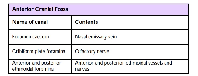
Name the foramina within the anterior cranial fossa and what structures pass through them.
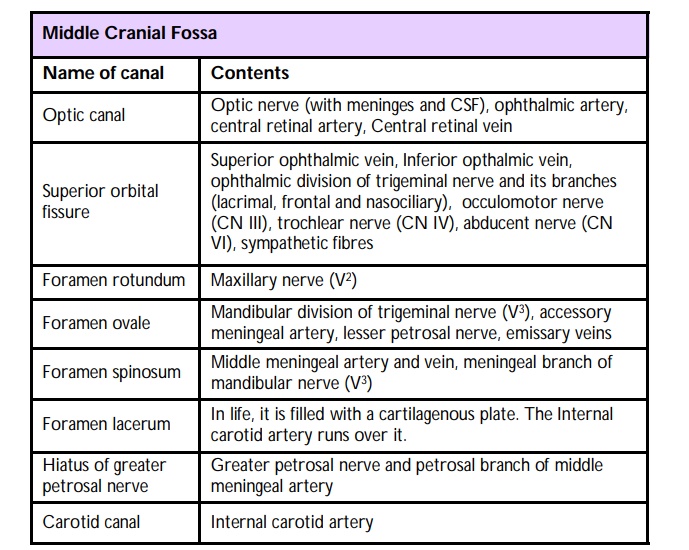
Name the foramina within the middle cranial fossa and what structures pass through them.
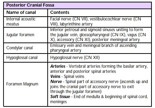
Name the foramina within the posterior cranial fossa and what structures pass through them.
Name the cranial sutures.
Metopic, Sagittal, Coronal(s), Lambdoid(s), Squamosal(s).
Describe why the suture lines are important in the context of suspected non-accidental injury in children.
"The persistent cranial sutures should not be mistaken for fractures during interpretation of X-rays or CT scans.
"
Which condition can result in early fusion of cranial sutures?
"Early fusion or premature closure of the sutures may cause craniosynostosis.
Note: Saggital synostosis is the most common form resulting in a ‘boat shaped’ skull.
"
What is a basal skull fracture?
"A basal skull fracture is a fracture that can involve one or more of the following bones: Temporal (commonest bone involved), occipital, sphenoid or ethmoid bones.
"
What are the signs and symptoms of a basal skull fracture?
"Signs of basal skull fracture include Battles sign (bruising of the mastoid process), Racoon eyes (bruising of the periorbital region). Symptoms include CSF
rhinorrhea, haematympanum, deafness and bleeding from the nose and ears, anosmia, facial nerve palsy.
"
What is the clinical relevance of the danger triangle of the face?
"The danger triangle of the face is the area from the corners of the mouth to the bridge of the nose, including the nose and maxilla. As there are no valves in the veins draining this area, retrograde infections from the nasal area can spread to the cranial cavity causing cavernous sinus thrombosis, meningitis or brain abscess.
"
What are the complications of cavernous sinus thrombosis?
Cavernous sinus thrombosis can cause symptoms related to both orbital and cranial nerve involvement:
• Orbital symptoms include periorbital oedema, ptosis, chemosis, proptosis and photophobia.
• Cranial nelve palsies (CNIII, CNIV, CNV1, CN V1 and CN V2 ) can occur, with the sixth nerve (abducent) to be the first cranial nerve to be commonly affected. This is because the abducent nerve lies in the middle of the cavernous since as opposed to the other nerves that lie along the lateral wall of the cavernous sinus.