What is the origin of the median nerve?
"The median nerve originates from the lateral and medial cords of the brachial plexus.
"
What is the root value of the median nerve?
"The median nerve receives a contribution from all roots of the brachial plexus (C5, C6, C7, C8 and T1).
"
Describe the course of the median nerve.
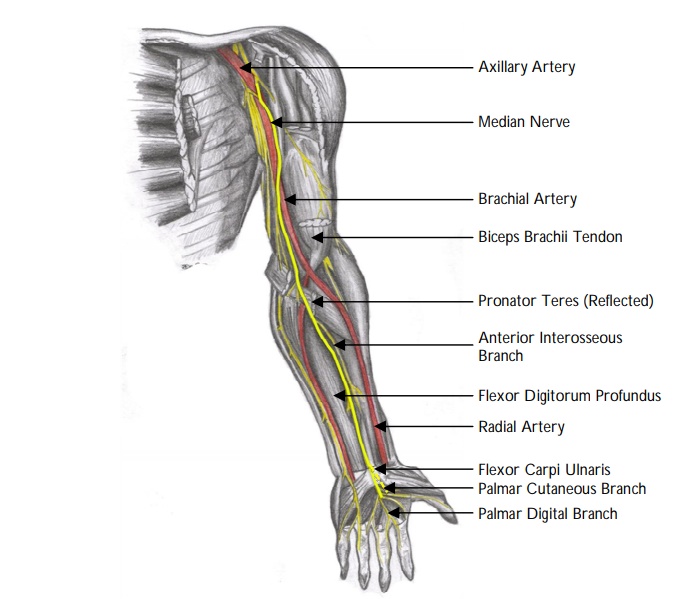
Formed in the axilla by the union of the lateral root from the lateral cord and the medial root from the medial cord of the brachial plexus.
It runs distally in the arm, initially on the lateral side of the brachial arteryuntil it reaches the middle of the arm, where it crosses from lateral to medial and lies over the brachialis.
Then descends to the cubital fossa, where it lies deep to the bicipital aponeurosis and median cubital vein and, medial to the brachial artery.
The median nerve does not give rise to any branches in the arm but supplies articular branches to the elbow joint.
Enters the forearm between the two heads of pronator teres where it gives rise to the anterior interosseous nerve.
Runs deep to flexor digitorum superficialis (FDS) until just proximal to the wrist where it becomes superficial to this tendon.
The median nerve passes through the wrist in the carpal tunnel entering between palmaris longus and flexor carpi radialis, superficial to flexor digitorum superficialis.
Palmar cutaneous nerve arises approximately 5 cm proximal to the distal wrist crease and runs over the flexor retinaculum to supply cutaneous innervation to the skin over the thenar eminence.
The origin of the recurrent motor branch is variable. It usually arises just distal to the flexor retinaculum.
Name the branches of the median nerve.
"
The median nerve gives off the:
- Anterior interosseous nerve.
- Palmar cutaneous branch of the median nerve.
- Recurrent motor branch of the median nerve.
- Articular branches to the elbow joint.
Name the muscles directly innervated by the median nerve in the forearm.
"
The superficial layer of forearm flexor muscles of the forearm are innervated directly by the median nerve:
- Pronator teres (PT)
- Flexor carpi radialis (FCR)
- Palmaris longus (PL)
- Flexor digitorum superficialis (FDS)
What is the origin of the anterior interosseous nerve?
"
The anterior interosseous nerve is a motor branch of the median nerve. It arises between the two heads of pronator teres and runs on the anterior surface of the interosseous membrane.
Note: The posterior interosseous nerve is a branch of the radial nerve. It passes between the two heads of supinator to enter the posterior compartment of the forearm. It innervates most of the extensors of the wrist and digits.
"
Name the muscles innervated by the anterior interosseous nerve.
"
The deep layer of forearm flexors are innervated by the anterior interosseous nerve:
- Radial half of flexor digitorum profundus (FDP).
- Flexor pollicis longus (FPL).
- Pronator quadratus.
Which structures in the flexor compartment of the forearm are not supplied by the median nerve?
In the forearm, the median nerve supplies the long flexors of the forearm, with the exception of flexor carpi ulnaris and ulnar half of flexor digitorum profundus (which are innervated by the ulnar nerve).
What is the origin of the anterior interosseous artery?
"The anterior and the posterior interosseous arteries are branches of the ulnar artery.
"
What is anterior interosseous nerve syndrome (AINS)?
"Anterior interosseous nerve syndrome is a nerve entrapment syndrome resulting in a loss of motor function in flexor pollicis longus, flexor digitorum profundus to index and middle fingers, and pronator quadratus, without any sensory deficit.
"
How would you clinically test for anterior interosseous nerve syndrome (AINS)?
"
- Typically, patients fail to make an ‘O.K. Sign’, as flexion of the interphalangeal joint of the thumb and the distal interphalangeal joint of the index finger are impaired.
- Another sensitive test is the pinch test due to impairment of pincer function due to weakness of flexion of the distal phalanges of the thumb and index finger. For example, patient with AINS will also not be able to pinch a sheet of paper between the thumb and index finger.
- Weakness of the pronator quadratus muscle – weak resistance to forced supination in the distal radioulnar joint (to test the function of pronator quadratus one must eliminate the rotatory action of the pronator teres by fully flexing the elbow).
- There is no sensory deficit as the AIN is purely a motor nerve.
What is the origin of the recurrent motor branch of the median nerve?
The recurrent motor branch of the median nerve is usually arises just distal to or within the carpal tunnel and then hooks back to supply the thenar muscles. However, it can also exit through or superficial to the flexor retinaculum and is therefore at risk of injury during carpal tunnel decompression.
What is the innervation of the muscles of the hand.
The ulnar nerve is the motor supply to all the intrinsic muscles of the hand except ‘LOAF’ which are supplied by the motor recurrent branch of the median
nerve:
Lateral (radial) two lumbricals
Opponens pollicis (OP)
Abductor pollicis brevis (APB)
Flexor pollicis brevis (FPB) (superficial head)
What is the sensory innervation of the hand?
"
- Median nerve:
- The palmar cutaneous branch of the median nerve arises approximately 5 cm proximal to the distal wrist crease, runs over the flexor retinaculum and supplies sensation to the thenar skin. The median nerve runs under the retinaculum, gives off the recurrent motor branch and the then supplies sensation to the radial three and a half digits.
- Ulnar nerve:
- The ulnar nerve supplies the sensation to the ulnar one and a half digits (the little finger and the ulnar half of the ring finger).
- The dorsal branch of the ulnar nerve arises approximately 5 cm proximal to the wrist and supplies the ulnar side of the little finger and dorsum of the ulnar 1½ digits.
- The ulnar nerve divides into a deep motor branch and continues as a superficial palmar branch as it passes through Guyon’s canal on the lateral side of the pisiform bone. The superficial palmar branch supplies the skin of the volar 1½ digits and hypothenar eminence respectively.
- Radial nerve:
- The superficial radial nerve supplies the sensation to the skin of the dorsum of the radial 3½ digits up to the level of the proximal phalanx.
"