Name the origin of the extensor muscles of the forearm.
"
- Common extensor origin (tendon that attaches to the lateral epicondyle of the humerus):
- Extensor carpi radialis brevis, extensor digitorum communis, extensor digiti minimi and extensor carpi ulnaris.
- Lateral supracondylar ridge of the humerus:
- Brachioradialis and extensor carpi radialis longus.
- Radius, ulna and interosseous membrane:
- Extensor indices, extensor pollicis longus, extensor pollicis brevis and abductor pollicis longus.
- Lateral epicondyle of the humerus:
- Anconeus.
- Supinator (also arises from the superior crest of the ulna, radial collateral ligament and the annular radial ligament).
What is the innervation of the muscles on the radial aspect of the forearm?
"
All the muscles on the radial (extensor) aspect of forearm (with the exception of anconeus, brachioradialis and extensor carpi radialis longus which are supplied by the radial nerve) are supplied by the posterior interosseous nerve (PIN):
- Extensor carpi radialis brevis.
- Extensor digitorum communis.
- Abductor pollicis longus.
- Extensor indices.
- Extensor pollicis longus
- Extensor pollicis brevis.
- Extensor carpi ulnaris.
- Extensor digiti minimi.
Name the boundaries of the anatomical snuff box.
"
- Ulnar border : Extensor pollicis longus (EPL) tendon.
- Radial border : Tendons of abductor pollicis longus (APL) and extensor pollicis brevis (EPB).
- Proximal border : Radial styloid process.
- Floor : Scaphoid and trapezium.
Name the contents of the anatomical snuffbox?
"
From superficial to deep the contents of the anatomical snuffbox are the:
- Cephalic vein
- Superficial terminal branches of the radial nerve.
- Extensor carpi radialis longus and brevis tendons lie on the ulnar side of the snuffbox.
- Radial artery.
- Bones (radial styloid, scaphoid, trapezium, base of the first metacarpal).
Describe the arrangement of the extensor tendon fascial compartments
I : Extensor pollicis brevis and abductor pollicis longus.
II : Extensor carpi radialis longus and extensor carpi radialis brevis.
III : Extensor pollicis longus.
IV : Extensor indicis and extensor digitorum communis.
V : Extensor digiti minimi.
VI : Extensor carpi ulnaris.
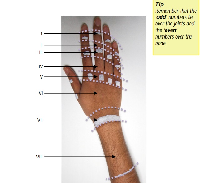
Describe the zones of the extensor tendon injuries.
"
- Zone I : Level of the DIPJs.
- Zone II : Level of the middle phalanges.
- Zone III : Level of the PIPJs.
- Zone IV : Level of the proximal phalanges.
- Zone V : Level of the MCPJs.
- Zone VI : Level of the metacarpals.
- Zone VII : Level of the wrist joint.
- Zone VIII : Distal forearm (at the level of the musculotendinous junction).
- Zone IX : Proximal forearm.
Describe the most common mechanism of a mallet finger injury.
"A zone I extensor tendon injury is known as mallet finger injury. This is due to division, or more commonly, avulsion of the extensor tendon apparatus at the DIPJ. The most common mechanism is forced hyper-flexion of the DIPJ such as when a ball or force strikes the tip of the finger or even in everyday activities such as tucking in bed sheets. It can also occur in forced hyperextension. The finger may be painful, swollen and bruised, especially if there is an associated fracture. However, often the only presentation is the inability to straighten the tip of the finger (i.e., the patient is unable to extend the finger at the DIPJ).
"
How would you manage a mallet finger injury?
"The majority of mallet injuries can be treated conservatively with a mallet splint for 6-8 weeks (to keep the DIPJ extended until healing is complete). Surgical repair may be necessary (dermatotenodesis/ open repair +/- K-wire) if there is a large bone fragment or poor alignment.
"
Describe the extensor tendon zones of the thumb.
"The zones of the thumb differ from the fingers as there are only two phalanges. They are numbered I-V with zones I overlying the IPJ, zone II over the proximal phalanx, zone III over the MCPJ, zone IV over the first metacarpal, and zone V over the wrist joint.
"
Name some causes of spontaneous rupture of the extensor pollicis longus tendon.
"
Spontaneous rupture of the extensor pollicis longus (EPL) tendon is associated with:
- Rheumatoid arthritis.
- Fractures of the wrist.
- Gout.
- Systemic or local steroids.
- Ankylosing spondylitis.
- Bony spurs from metastases.
- Lister’s tubercle deformities.
- Repetitive movement of the wrist joint.
"
Describe the mechanism of a Boutonnière Deformity.
"Rupture or avulsion of the central slip leads to Boutonnière deformity due to loss of PIPJ extension and later DIPJ hyperextension.
"
Briefly describe some of the common hand deformities seen in rheumatoid arthritis.
Ulnar deviation of the wrist : Volar subluxation of the carpus from the ulna.
Subluxation : Volar and ulnar subluxation of the MCPJs.
Swan neck deformity : Hypertension at the PIPJ and flexion at the DIPJ.
Boutonnière deformity : Flexion at the PIPJ and extension at the DIPJ.
‘Z’- shaped thumb : Flexed CMCJ and IPJ with extended (similar to swan neck deformity).
What are the radiological features of rheumatoid arthritis?
The radiological features of RA are:
Joint space narrowing.
Joint erosions.
Periarticular cysts.
Soft tissue swelling.