THORAX
OSCE
Station 10
The Lungs and Trachea
Describe the lobar arrangement of the lungs.
"
- The right lung has three lobes; upper, middle and lower, divided by the oblique and horizontal fissures (horizontal divides upper and middle, oblique divides upper and middle from lower).
- The left lung has only an upper and lower lobe, divided by the oblique fissure.
- The counterpart of the middle lobe of the right lung in the left lung is the lingula, which lies between the oblique fissure and cardiac notch.
What is a bronchopulmonary segment?
"A segment of lung consisting of a segmental/tertiary bronchus, a segmental branch of the tertiary arteries, a segment of the lung tissue, and the surrounding connective-tissue septum.
"
How many bronchopulmonary segments does each lung have?
"
- The right lung has 10 bronchopulmonary segments. The upper lobe has three segments, the middle lobe has two segments and the lower lobe has five segments.
- The left lung has eight bronchopulmonary segments. The upper and lower lobes both have four segments each (therefore eight segments in total).
Describe the anatomy of a bronchopulmonary segment.
"
Each bronchopulmonary segment is:
- Pyramidal shaped with the apices facing the lung root and the base facing the pleural surface.
- Supplied by its own tertiary arteries (from the pulmonary and bronchial arteries, and run together through the centre of the segment).
- It is separated from the surrounding lung by a layer of connective tissue.
- Veins and lymphatic vessels drain along the edges of the segment.
What is the clinical significance of the bronchopulmonary segments?
"Each bronchopulmonary segment is a discrete anatomical and functional unit, and this separation means that a bronchopulmonary segment can be surgically removed without affecting the function of the others.
"
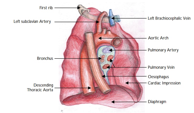
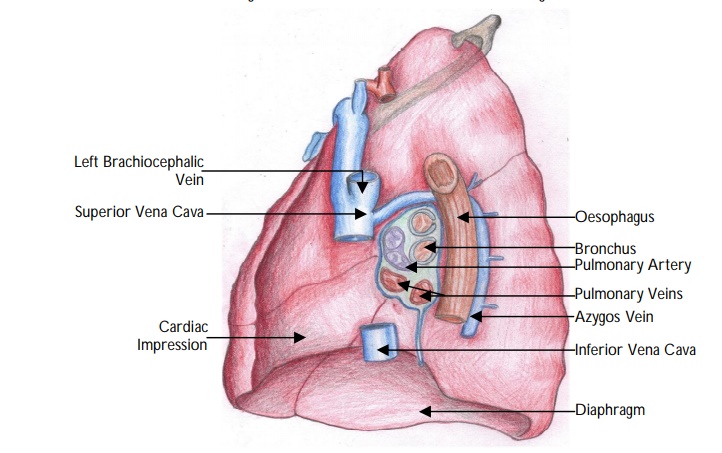
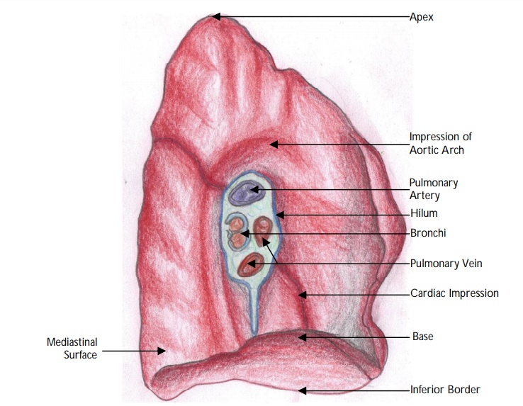
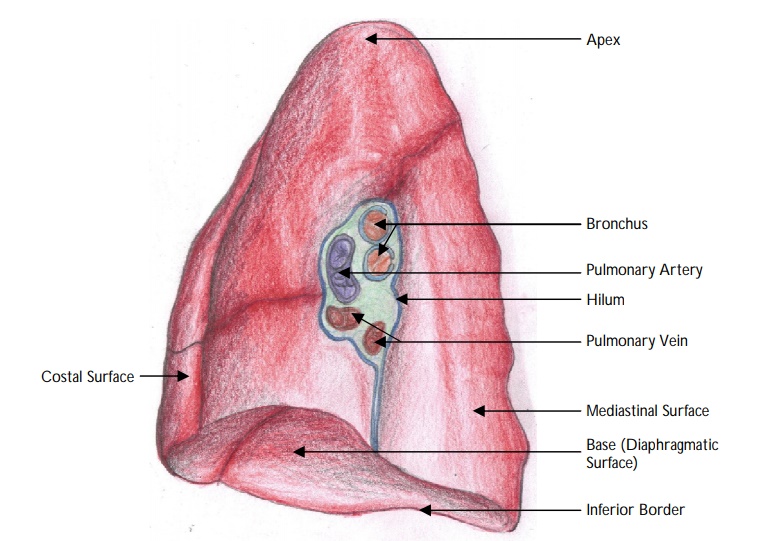
Label the structures on the two images below:
View answer
Describe the anatomy of the trachea.
"
- The trachea commences at the lower border of the larynx (cricoid cartilage) at the level of C6 vertebrae and measures approximately 10-15 cm in length.
- The cricoid cartilage is the only complete ring of cartilage in the trachea. Below that there are 15-20 incomplete C-shaped tracheal rings of cartilage, which re-inforce the front and sides of the trachea.
- The trachealis muscle joins the ends of the incomplete rings and are connected by bands of fibrous connective tissue known as the annular ligaments of the trachea.
- The posterior surface has a membranous wall devoid of cartilage.
- The incomplete rings allow the trachea to contract during coughing and collapse during swallowing (to allow the oesophagus to accommodate food).
- Bifurcates into right and left main bronchi at the level of the sternal angle (T4/T5 vertebral level).
- The carina is situated to the left of the median line and the right bronchus is usually a direct continuation of the trachea than the left.
Describe the important anatomic relations of the trachea in the neck.
"
The important anatomic relations of the trachea in the neck are:
Anterior:
- Isthmus of the thyroid gland.
- Inferior thyroid veins.
- Thyroidea ima artery (if present).
- Sternothyroid and sternohyoid muscles.
- Cervical fascia.
- Jugular venous arch.
Lateral:
- Common carotid arteries.
- Right and left lobes of the thyroid gland.
- Inferior thyroid arteries.
- Recurrent laryngeal nerves (postero-lateral).
Posterior:
- Oesophagus.
- Recurrent laryngeal nerves (lying in the tracheo-oesophageal groove).
- Vertebral column.
Describe the important anatomic relations of the trachea in the thorax.
"
In the thorax, the trachea lies in the superior mediastinum. Its anatomic relations in the thorax are:
Anterior:
- Manubrium sterni.
- Remnants of the thymus.
- Left brachiocephalic vein.
- Aortic arch.
- Brachiocephalic trunk.
- Left common carotid artery.
- Deep cardiac plexus.
Lateral:
- Right side : Vagus nerve, azygos vein, pleura and, near the root of the neck with the brachiocephalic trunk.
- Left side : Aortic arch, left common carotid artery, left subclavian arteries, left recurrent laryngeal nerve, pleura.
Posterior:
- Oesophagus.
- Recurrent laryngeal nerves (lying in the tracheo-oesophageal groove).
Describe the course of the right main bronchus.
"
The right main bronchus:
- Is approximately 2.5 cm in length.
- It is wider and shorter with a more vertical course than the left.
- Takes an inferior and lateral course behind the ascending aorta and SVC to enter the lung hilum.
- Gives off the upper lobe bronchus before entering the lung, then divides into the bronchi to the middle and inferior lobes within the lung.
Describe the course of the left main bronchus.
"
- Is approximately 5 cm in length.
- Passes below the arch of the aorta, anterior to the oesophagus and descending aorta.
- The pulmonary artery first lies anterior, then superior to the bronchus.
- The left main bronchus gives off no branches before entering the hilum of the lung, where it divides into the bronchi to upper and lower lobes.
What layers would you pass through pass through when performing a tracheostomy?
Skin (a vertical or transverse skin incision can be used).
Superficial cervical fascia (and platysma laterally in the transverse incision).
Pretracheal fascia.
Note: the isthmus of the thyroid may be encountered, which will need to be retracted
superiorly or divided to expose the trachea
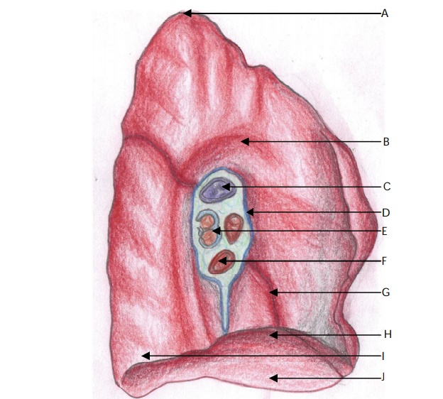
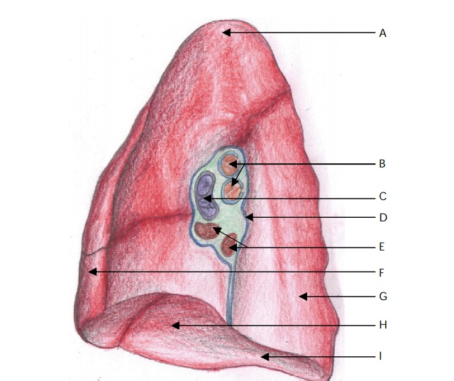
Label the parts on the images below:
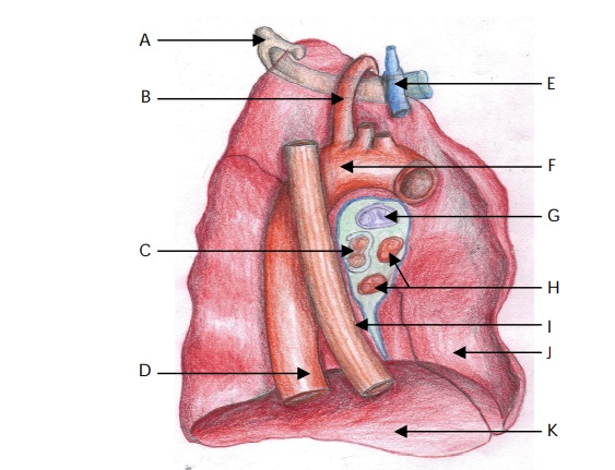
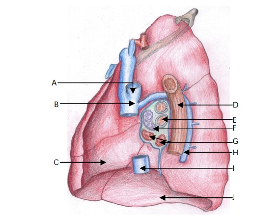
View answer