Describe the blood supply to the foot.
"
- The blood supply to the foot is mainly derived from the posterior tibial artery.
- The posterior tibial artery terminates by dividing into the medial and lateral plantar arteries (under the flexor retinaculum).
- The medial plantar artery passes along medial aspect of the sole of the foot and terminates as digital branches which join with the plantar metatarsal branches of the plantar arch.
- The lateral plantar artery passes obliquely across the sole of the foot giving off muscular and cutaneous branches.
- It anastomoses with the dorsalis pedis artery to form the plantar arch, which give rise to the metatarsal arteries which, in turn give rise to the plantar digital arteries which contribute to the blood supply of the toes.
Name the structures which pass behind the medial malleolus.
"
The structures which pass behind the medial malleolus from anterior to posterior are:
- Tibialis posterior.
- Flexor Digitorum longus.
- Tibial Artery.
- Venae Comitantes.
- Tibial Nerve.
- Flexor Hallucis longus.
Name the tarsal bones.
"
The tarsal bones, from proximal to distal are:
- Proximal : Talus and Calcaneus.
- Intermediate : Navicular.
- Distal : Cuboid and three Cuneiforms (lateral, intermediate and medial).
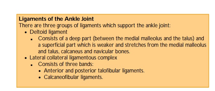
Name the three groups of ligaments of the ankle joint.
"
The three groups of ligaments which support the ankle joint are the:
- Deltoid ligament.
- Lateral collateral ligamentous complex.
- Syndesmosis.
"
Name the arches of the foot.
"
The arches of the foot are the:
- Medial longitudinal arch.
- Lateral longitudinal arch.
- Transverse arch (each foot contributes half of the transverse arch).
Which ligament supplies the main support of the medial longitudinal arch of the foot?
| The plantar calcaneonavicular ligament (spring ligament) connects the sustentaculum tali with the plantar surface of the navicular bone. It provides the main support for the medial longitudinal arch of the foot. |
Describe the landmarks where you would palpate the pulses in the foot and ankle.
"
- The landmarks for palpation of the Dorsalis pedis artery are between the tendons of extensor hallucis longus and extensor digitorum on the dorsum of the foot, lateral to the base of the first metatarsal.
- Any one of the following landmarks can be used for palpation of the
Posterior tibial artery:
1) 2 cms below and behind the medial malleolus; 2) one third of the way between the medial malleolus and calcaneum; 3) half-way between the medial malleolus and the Achilles tendon.
"
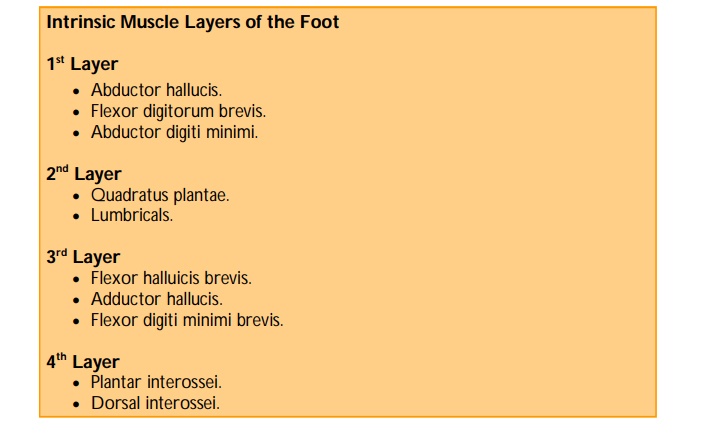
How many intrinsic layers of muscles are there on the plantar aspect of the foot?
| There are four layers of intrinsic muscles on the sole of the foot. |
Name the muscles that make up the first layer of the plantar aspect of the foot.
"
The muscles that make up the first layer of the foot are the:
- Abductor hallucis.
- Flexor digitorum brevis.
- Abductor digiti minimi.
Describe the function of the dorsal and plantar interossei of the foot.
"
- The plantar interossei adduct the digits towards the second digit.
- The dorsal interossei abduct the digits away from the second digit. Together with the lumbricals they extend the DIPJs, PIPJs and flex the MTPJs.
"
Name the nerve which innervates the dorsal and plantar interossei.
The dorsal and plantar interossei are innervated by the lateral plantar nerve.
Name the cutaneous innervation of the medial aspect of the foot.
The saphenous nerve supplies skin on the medial aspect of the foot (anteriorly to the head of the first metatarsal).
Note: The sural nerve supplies sensation to the lateral aspect of the foot.
How are ankle fractures classified?
"
Ankle fractures are classified using the Weber classification:
- A : Fracture is below the distal talo-fibular joint (a syndesmosis).
- B : Fracture is at the level of the syndesmosis.
- C : Fracture is above the level of the syndesmosis.
What is the clinical relevance of the Weber classification?
| Fractures at or above the level of the syndesmosis (Weber B and C) are likely to produce an unstable ankle joint and are therefore more likely to require open reduction and internal fixation Weber A fractures can be managed conservatively with PoP cast or operatively with AO screws. |
What is a stress fracture?
| A stress fracture is an incomplete fracture caused by repeated stress and occurs most frequently to metatarsals II, III and IV. It is common in soldiers and athletes. |
What is tarsal tunnel syndrome?
"
Tarsal tunnel syndrome may be described as a constellation of signs and symptoms (usually pain and paraesthesia) caused by entrapment or compression of the tibial nerve or any of its branches in the region beneath the flexor retinaculum in the medial aspect of the ankle. Causative factors include obesity, pes planus, repetitive strain and any compressive lesions. Tarsal tunnel syndrome may refer to either ‘anterior’ or ‘posterior’ tarsal tunnel syndrome:
- Anterior tarsal tunnel syndrome is entrapment of the terminal portion of the deep peroneal nerve as it runs below the dense superficial fascia of the ankle. It is a rare neuropathy and results in pain and paraesthesia over the 1st dorsal webspace.
- Posterior tarsal tunnel syndrome results from entrapment of the posterior tibial nerve at the level of the medial maleolus resulting in pain and paraesthesia of the sole of the foot. It is also uncommon.
How would you diagnose tarsal tunnel syndrome.
| The diagnosis is usually made clinically by palpating along the clinical course of the nerve in the tarsal tunnel or by percussion of the nerve (Tinnel’s test) to elicit discomfort either locally or distally. Ultrasound or MRI may be useful in investigating the causative pathology. |
What is the management of tarsal tunnel syndrome?
"
- Conservative management : Rest, elevation, NSAIDs, analgesia (including neuropathic agents), supportive footwear/orthoses.
- Minimally invasive : Local anaesthetic or corticosteroid injections
- Surgical : Surgical excision of any compressive lesion or tarsal tunnel release.