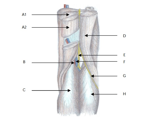
Identify the points on this image
View Answer
Name the boundaries of the popliteal fossa.
"
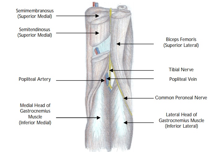
The boundaries of the popliteal fossa are:
- Superior medial : Semimembranosus and semitendinosus.
- Superior lateral : Biceps femoris.
- Inferior medial : Medial head of gastrocnemius.
- Inferior lateral : Lateral head of gastrocnemius.
- Roof : Popliteal fascia, subcutaneous tissue, skin.
- Floor : Popliteal surface of femur, capsule of the knee joint and popliteus.
Name the contents of the popliteal fossa.
"
The contents of the popliteal fossa from superficial to deep are the:
- Common peroneal nerve.
- Tibial nerve.
- Popliteal vein.
- Popliteal artery.
- Capsule of the knee joint.
- Tendon of popliteus laterally.
Note: Superiorly, the popliteal fossa contains the sciatic nerve only (before it divides).
The popliteal fossa also contains:
- Termination of the small saphenous vein as it drains into the popliteal vein.
- Sural nerve (arising from the tibial nerve).
- Five genicular branches of the popliteal artery,
- Lymph nodes.
- Fat.
What is the clinical significance of this anatomy when performing peroneal nerve block?
In peroneal nerve blocks, the common peroneal nerve is located most superficial of all the structures, followed by the popliteal vein, then the popliteal artery which is the deepest structure from the needle.
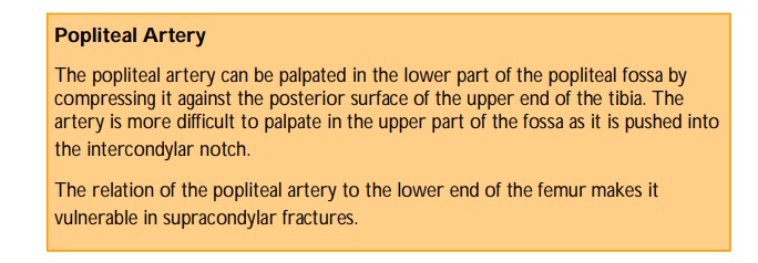
Where does the popliteal artery commence?
"The popliteal artery is a continuation of the femoral artery. It begins at the adductor hiatus and runs infero-laterally.
"
Describe the blood supply to the knee joint.
"
- Five genicular branches of the popliteal artery supply the articular capsule and ligaments of the knee joint.
- The genicular arteries are the lateral superior, medial superior, middle, lateral inferior, and medial inferior genicular arteries.
- They form a genicular anastomoses, a network of vessels, around the knee joint.
- Other vessels contribute to this anastomosis:
Descending genicular branch of the femoral artery.
Descending branch of the lateral femoral circumflex artery (superolaterally).
Anterior recurrent branch of the anterior tibial artery (inferolaterally).
Describe the course of the popliteal artery as it exits the popliteal fossa.
"
- The popliteal artery usually bifurcates as it exits the popliteal fossa in 80-90% of the population but it can trifurcate in a small proportion of individuals.
- When the artery bifurcates it divides into an anterior tibial artery and tibio-peroneal trunk which then divides into the posterior tibial and peroneal (fibular) arteries.
- When the artery trifurcates it divides into: anterior tibial, posterior tibial and peroneal (fibular) arteries.
- In addition to the genicular branches and the main vessels described above, the sural arteries (inferior muscular branches) also arise from the popliteal artery which supply the gastrocnemius, soleus and plantaris.
- The superior muscular branches of the popliteal artery have clinically important anastomoses with the terminal part of the deep femoral and gluteal arteries.
Describe the clinical course of the common peroneal nerve.
"
- The common peroneal (fibular) nerve leaves the fossa by passing superficial to the lateral head of gastrocnemius and biceps femoris then passes over the posterior aspect of head of the fibula.
- The nerve then then winds around the neck of the fibula deep to peroneus longus.
- It then descends and divides into superficial and deep peroneal branches .
What does the superficial peroneal nerve supply?
The superficial peroneal nerve supplies the lateral compartment of the leg and the skin over the 2nd - 4th webspaces over the dorsum of the foot.
Describe why the common peroneal nerve is vulnerable to injury.
"
- The common peroneal nerve is susceptible to injury from trauma since it is quite superficial as it winds around the lateral part of the neck of the fibula.
- It can be injured at this site in adduction injuries to the knee or compressed by a tight plaster cast or firm bandage.
"
What is the clinical significance of injury to this nerve?
This results in paralysis of the dorsiflexors and the patient presents with footdrop. In addition, the eversion of the foot is lost and the inversion is weak.
What are the differential diagnoses of a swelling in the popliteal fossa?
The differential diagnoses of a swelling in the popliteal fossa include:
Baker's cyst (also known as a popliteal cyst, is a benign swelling in the posterior aspect of the knee, associated with rheumatoid arthritis).
Popliteal artery aneurysm
Popliteal abscess
Neoplasm
Varicose veins
Deep vein thrombosis
Lymphadenopathy
Lipoma
Which cutaneous dermatome supplies sensation over the anterior and posterior aspects of the knee knee?
"
- Anterior : L3 dermatome.
- Posterior : S1, S2.