Describe the blood supply to the liver.
"
The liver receives blood from two sources: the portal vein (70-80%) and hepatic artery (20-30%)
- Hepatic artery supplies oxygen-rich blood from the aorta.
- Blood from the portal vein is oxygen poor but nutrient rich carried from the gastrointestinal tract to the liver sinusoids.
Describe the vascular segments of the liver.
"
- The liver is divided into eight vascular segments, based on the divisions of the hepatic artery, portal vein and hepatic ducts.
- Each segment of the liver receives a branch of the hepatic artery and portal vein, and is drained by a branch of the bile duct.
- The intersegmental hepatic veins run between the segments.
- Segments 1-4 are considered to be the left lobe of the liver and segments 5-8 are considered to be the right lobe.
What is the function of the hepatic veins?
"The hepatic veins of the liver run between the segments, draining adjacent segments. The hepatic veins drain into the inferior vena cava just below the diaphragm.
"
Why are the vascular segments of the liver clinically significant?
The arrangement of vascular segments of the liver makes it possible to perform hepatic lobectomies without excessive bleeding, and more recently to remove only the affected segment (‘segmentectomies’) for example, in malignancy.
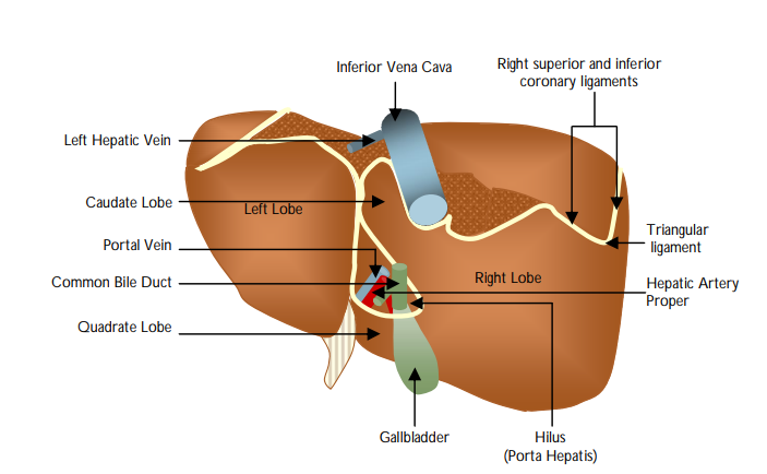
Describe the ligaments of the liver.
"
- Falciform ligament: A reflection of peritoneum extending between the upper anterior abdominal wall and the liver. It encloses the round ligament of the liver in its inferior free edge.
- Round ligament of the liver: This is the fibrosed remnant of the umbilical vein that carried well-oxygenated and nutrient rich blood from the placenta to the foetus. The umbilical vein joins the ligamentum venosum (the ductus venosus in the foetus). In utero, the ductus venosus allows umbilical blood from the placenta to bypass the liver and drain directly into the IVC and pass to the foetal heart.
- Coronary ligament: This is formed from reflections of peritoneum onto the diaphragmatic surface of the right and left lobes of the liver, meeting on the right to form the right triangular ligament. The coronary ligament has anterior and posterior layers. The anterior layer of the coronary ligament is continuous with the falciform ligament. The posterior layer is continuous with the lesser omentum.
What is the bare area of the liver?
"The bare area of the liver is a triangular part of the liver not covered by visceral peritoneum because it is in direct contact with the diaphragm. It is located between the layers of the coronary ligament.
"
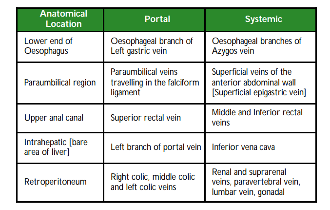
What are porto-systemic anastomosis?
"Porto-systemic anastomoses are communications between the portal venous system and the systemic venous system. Normally, these communications are not patent; however, in portal hypertension these channels open up leading to engorgement of the veins and subsequent bleeding.
"
Name some anatomical sites of porto-systemic anastomoses
What is the management of acute oesophageal variceal bleeding?
"
- The patient should be resuscitated as per ALS guidelines.
- Terlipressin is recommended for all patients with suspected variceal bleeding at presentation.
- Endoscopic banding.
- Transjugular intrahepatic portosystemic shunts (TIPS) can be considered if banding fails.
- Balloon tamponade with Sengstaken-Blakemore tube is not in the current guidelines but can be used to temporise either prior to endoscopy (if immediate endoscopy is not available) or between failed banding and TIPS.