THORAX
OSCE
Station 5
The Heart
What is the first branch of the ascending aorta?
"The coronary arteries are the first branch of the ascending aorta.
"
What is the origin of the right coronary artery?
"The right coronary artery arises from the right aortic sinus of the ascending aorta.
"
What territory does the right coronary artery supply?
"
The right coronary artery supplies:
- All surfaces of right atrium.
- About 25-30% of right ventricle.
- The posterior descending artery that arises from the right coronary in majority of individuals, supplies the inferior wall of right ventricle, ventricular septum, and the posteromedial papillary muscle.
- Sinoatrial (SA) node (in 60% of people) and atrioventricular (AV) node (in majority of individuals).
What is the origin of the left coronary artery?
"The left coronary artery arises from left aortic sinus of the ascending aorta.
"
What territory does the left coronary artery supply?
"
The left coronary artery supplies the:
- Left atrium.
- Most of the left ventricle.
- Part of the right ventricle.
- Most of the interventricular septum.
- SA node in 40%.
Which coronary arteries are most commonly occluded?
"
The most common sites of coronary artery occlusion are the:
- Anterior branch of the left coronary artery (40-50%).
- Right coronary artery (30-40%).
- Circumflex branch (15-20%).
Name the conduction pathways in the heart.
"
- The sino-atrial (SA) node.
- The atrio-ventricular (AV) node.
- The bundle of His.
- The left and right bundle branches.
- The Purkinje fibres.
What is the blood supply to the conduction system?
"
- SA node is supplied by the SA nodal artery that arises from the right coronary artery in 60% and the left coronary artery in 40% of individuals.
- The AV node is supplied by the AV nodal branch that arises from the right coronary artery.
- The right bundle branch is supplied by the left coronary artery.
- The left bundle branch is supplied by the right and left coronary arteries.
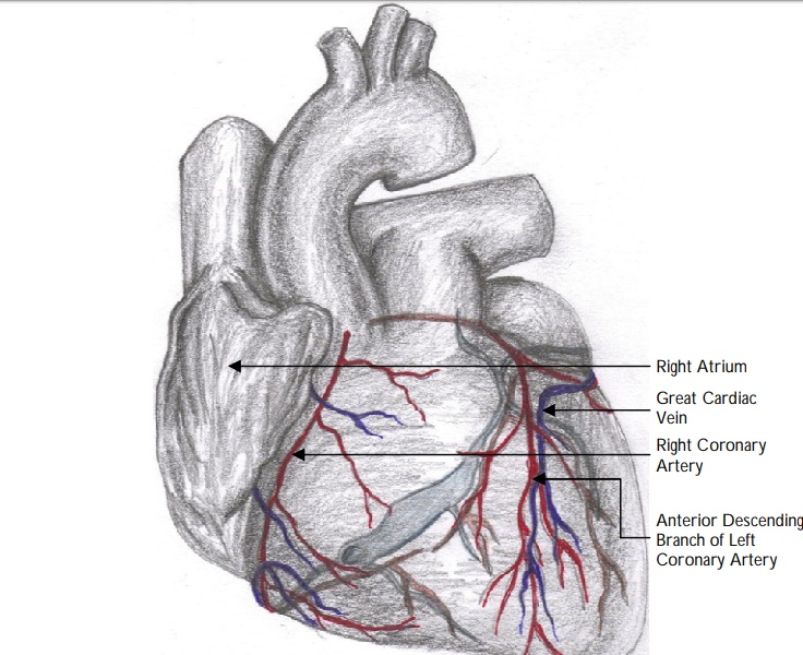
Identify the structures on the image
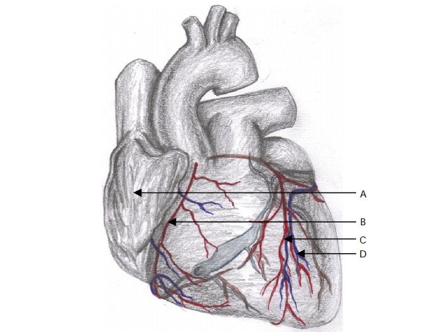
View answer
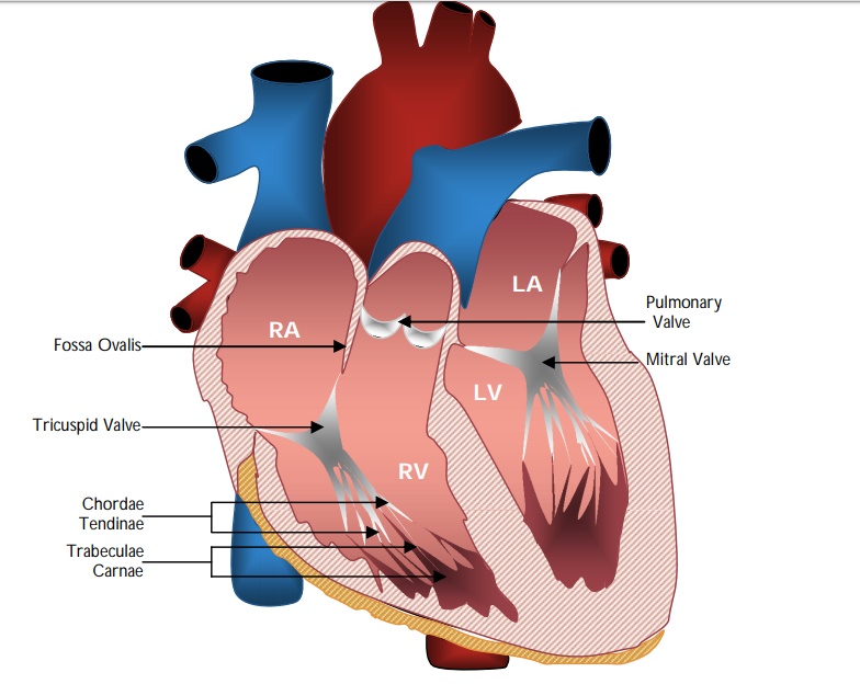
Identify the structures on the image:

View answer
What is the function of the chordae tendinae?
"The chordae tendinae are cord-like tendons that connect the papillary muscles to the tricuspid and mitral valves. They prevent eversion of the leaflets (i.e., cusps of the bicuspid and tricuspid valves) into the atrium during ventricular contraction.
"
What is the bundle of His and what is its function?
The bundle of His (also known as atrioventricular bundle) are a group of specialised cardiac muscle cells that conducts electrical impulses from the atrioventricular node in the right atrium to the septum between the ventricles and then to the left and right ventricles. Initially, it branches into left and the right bundle branches, which run along the interventricular septum. The left bundle branch further divides into left anterior and left posterior fascicles. These bundles and fascicles give rise to Purkinje fibers which distribute the impulse to the ventricular muscle. It takes about 0.03–0.04 seconds for the impulse to travel from the bundle of His to the ventricular muscle.
Describe the anatomy and function of the pericardium.
The pericardium, a double-walled sac containing the heart and the roots of the great vessels, has two layers – an outer fibrous layer and an inner serous layer.
The inner serous layer is further divided into visceral and parietal layers.
The visceral pericardium is part of the epicardium and the parietal pericardium is fused to the fibrous pericardium.
The pericardial cavity is a potential space between the visceral and parietal layers, and contains the pericardial fluid, that helps in lubricating the heart to prevent friction during cardiac activity.
The fibrous pericardium fuses with the adventitia of the great vessels at its root (i.e., the origin).
At the base, the fibrous pericardium becomes continuous with the central tendon of the diaphragm.
The pericardium, in general, helps to fix the heart in the mediastinum, protects the heart from infections, lubricates the heart and prevents excessive dilation of the heart (specifically by the outer tough fibrous layer) in cases of acute volume overload.