Describe the anatomy of the cervical plexus.
"
- The cervical plexus is a plexus of the first four cervical spinal nerves located from C1 to C4 in the neck.
- They are located laterally to the transverse processes and emerge from the posterior triangle midway on the posterior border of the sternocleidomastoid.
- They anastomose with the accessory nerve, hypoglossal nerve and sympathetic trunk.
- The cervical plexus has two types of branches: cutaneous and muscular.
Name the four cutaneous branches of cervical plexus and their innervation.
"
-
The four cutaneous branches of the cervical plexus are the:
- Lesser occipital nerve (C2 only) : Innervates lateral part of occipital region.
- Great auricular nerve (C2, C3) : Innervates skin near concha, auricle, external acoustic meatus, parotid region and post auricular region.
- Transverse cervical nerve (C2, C3) : Innervates anterior region of neck.
- Supraclavicular nerves (C3, C4) : Innervates the supraspinatus, shoulder, and upper thoracic regions.
Name the four muscular branches of the cervical plexus and their innervations.
"
The four muscular branches of the cervical plexus are the:
- Ansa cervicalis (C1-C3) : Lies superficial to the internal jugular vein. Innervates sternohyoid, sternothyroid and omoyhoid muscles.
- Phrenic (C3-C5, primarily C4) : Innervates diaphragm and the pericardium.
- Communicating branches (C1) : Supplies geniohyoid and thyrohyoid muscles.
- Segmental branches (C1-C4) : Supplies anterior and middle scalene muscles.
"
Name the suprahyoid and infrahyoid strap muscles in the neck.
"
Suprahyoid muscles:
- Anterior belly of digastric
- Posterior belly of digastric
- Geniohyoid
- Stylohyoid
- Mylohyoid
Infrahyoid muscles:
- Superior belly of omohyoid
- Inferior belly of omohyoid
- Sternohyoid
- Sternothryroid
- Thyrohyoid
"
What is the nerve supply to the digastric muscle?
"
- The anterior belly of digastric is supplied by the nerve to mylohyoid, which is a branch of the mandibular division of the trigeminal nerve.
- The posterior belly if digastric is supplied by the digastric branch of the facial nerve.
What is Erb’s Point?
"Erb’s point is a point in the neck half way along the posterior border of the sternocleidomastoid muscle, from which the cutaneous nerves of the cervical plexus exit to become superficial to supply the skin. These nerves include the lesser occipital nerve, great auricular nerve, transverse cervical nerve, and supraclavicular nerve. In addition, the branches of the suprascapular nerve and the nerve to subclavius (from the upper trunk of the brachial plexus) lie deep to this point.
"
What is the clinical significance of Erb’s point?
"
A cervical plexus block can be achieved by infiltrating local anaesthesia at Erb’s point.
Note: Regional anaesthesia can produce an effective block for procedures involving neck, occipital region, shoulder and upper pectoral region.
"
What is the most likely diagnosis in a child presenting with a small pit on the anterior neck present since birth and having a recurrent mucinous discharge?
"
Right Second branchial cleft sinus/fistula.
The second branchial cleft anomalies accounts for majority of branchial anomalies (up to 90%). They are most frequently identified along the anterior border of the sternocleidomastoid muscle (junction of middle and lower 1/3rd).
"
What is the likely anatomical course of this?
"The second branchial fistula runs from the skin of the lateral neck, pierces platysma, ascends between the internal and external carotid arteries (close relation to the hypoglossal and glossopharyngeal nerve) and opens into the oropharynx, usually the palatine tonsil.
"
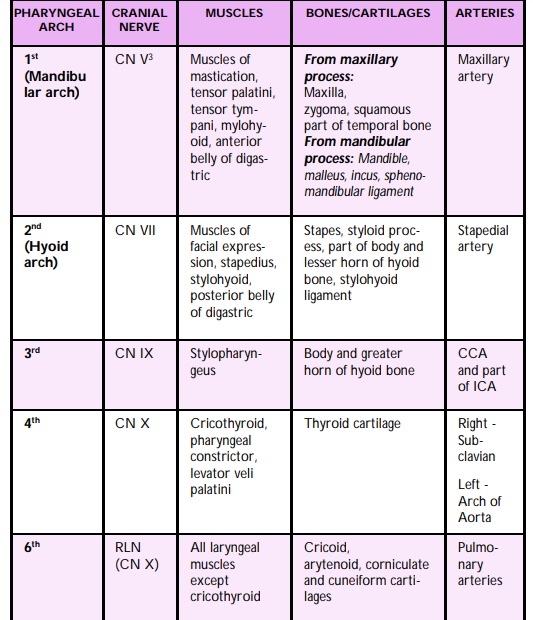
Describe the nerves, muscles and bony structures of the six pharyngeal arches